| Posted: Nov 18, 2014 |
Scientists X-ray nanoscale organelles of bacteria
|
|
(Nanowerk News) An international team of scientists led by Uppsala University has developed a high-throughput method of imaging biological particles using an X-ray laser. Images obtained with this method show projections of the carboxysome particle, a delicate and tiny cell organelle of photosynthetic bacteria. Organelles are tiny structures within the cell responsible for performing specific functions, much as organs within the body are responsible for performing certain functions.
|
|
This experiment, described in a paper published in the scientific journal Nature Photonics ("High-throughput imaging of heterogeneous cell organelles with an X-ray laser"), represents a major milestone for studies of individual biological structures using X-ray lasers like the European XFEL that is currently being built from the DESY campus in Hamburg to the neighbouring town of Schenefeld.
"The technique paves the way for 3D imaging of parts of the cell, and even small viruses, to develop a deeper understanding of life’s machinery", says Uppsala University professor Janos Hajdu, who is one of the lead authors on the paper and an advisor to European XFEL.
|
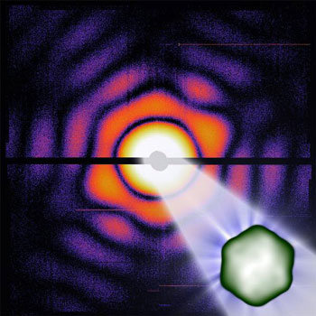 |
| Illustration of a carboxysome in the X-ray laser. (Image: Max Hantke/University of Uppsala)
|
|
To test the method, scientists from Uppsala University, the European XFEL, DESY and a number of other institutions studied the carboxysome, the cell organelle for carbon dioxide assimilation in cyanobacteria. Carboxysomes are responsible for about a third of global carbon fixation. The carboxysome contains protein machinery that incorporates carbon from carbon dioxide into biomolecules and has been studied extensively in Uppsala by Dirk Hasse and Inger Andersson. The carboxysome is a tiny icosahedral structure (a structure with 20 triangle-shaped sides) — of about 100 nanometres in diameter, too small to clearly see with an optical microscope.
|
|
Using a specially designed injector that produces a particle stream smaller than the width of a hair, the scientists sprayed an aerosol of carboxysomes across the beam of the LCLS X-ray laser at the SLAC National Accelerator Laboratory in the US.
|
|
“The structure of the organelles is determined from the way in which individual carboxysomes scatter the extremely short and ultra bright X-ray flashes of the LCLS”, says DESY scientist Anton Barty, one of the authors of the paper. Uniquely, this new method does not require crystals to get sufficient signal. “Thanks to the extreme brightness of the X-ray laser, which provides X-ray pulses of short enough duration to capture information before the sample explodes, we can reconstruct individual samples without having to crystallise the sample.”
|
|
Carboxysomes, like many other biological samples, cannot be crystallised.
|
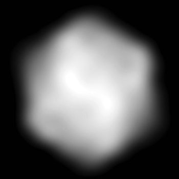 |
| Reconstructed image of an individual carboxysome. (Image: Max Hantke/University of Uppsala)
|
|
Within 12 minutes, approximately 70,000 scattering patterns were collected from individual particles. The analysis, using software developed in collaboration between Uppsala and DESY, returned an icosahedral shape for carboxysomes, in line with expectations. The results also showed considerable variation in size, ranging from 100 to 130 nm. This variability is what kept researchers from crystallizing the carboxysome.
|
|
"Our method allows single-particle imaging of objects which can be different in size and shape, which usually poses another challenge for experimenters”, says Max Hantke, a doctoral student in molecular biophysics at Uppsala University in Sweden who led the research. “Additionally, the size distribution of the carboxysomes before the experiment and what was seen in the resulting data matched almost perfectly”, Hantke says.
|
|
This suggests that the injection method did not cause the fragile carboxysomes to dissociate or clump together during their flight into the X-ray beam.
|
|
While high-resolution electron microscopy usually requires samples to be frozen, X-ray lasers like the LCLS or the European XFEL can analyse biological samples without freezing. This method also offers the possibility to image whole living cells at unprecedented resolution.
|
|
"With the carboxysomes we have reconstructed the smallest single non-crystallized biological particles ever imaged with an X-ray laser, and we also achieved the highest resolution for a biological particle imaged by the same method", explains Hantke. The reconstruction shows details as small as about 18 nanometres. "For the first time we access a very interesting size regime with an X-ray laser. Large pathogenic viruses like HIV, influenza, and herpes virus are in the same size domain as the carboxysome", says Hantke. “We hope this research could lead to three-dimensional models showing the diversity of nanoscale cell parts, which conventional techniques cannot access.”
|
|
“These advances lay the foundations for accurate, high-throughput structure determination by flash-diffractive imaging and offer a means to study structure and structural heterogeneity in biology and elsewhere”, says Hajdu. “In biology, there is heterogeneity and variability at all levels, and we needed a method for seeing it below the cellular level”, Hajdu says.
|
|
While the intense X-ray pulse destroys the sample, an accurate diffraction pattern can be acquired before it disintegrates. This method, called “diffraction-before-destruction”, was proposed in 2000 and demonstrated with non-biological samples at DESY’s FLASH facility in 2006.
|
|
Such single-particle imaging will be possible at the European XFEL when the facility opens to users in 2017. “With its 27,000 X-ray flashes per second and its high intensity, the European XFEL will open up more opportunities and possibilities for researchers”, Hajdu says. The dependence of structural variability on factors such as environment and genetics is of particular interest, and such studies require high throughput analysis methods to acquire sufficient statistics.
|
|
“These results show the way to high-throughput imaging of biological samples at high resolution”, says Filipe Maia, Hantke’s supervisor. “High data rates and very short X-ray flashes allow studies on the dynamics of particles and permit the analysis of structural variations, which are crucially important for life.”
|

