| Posted: Nov 10, 2015 |
Microwave field imaging using diamond and vapor cells
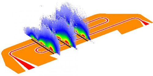
(Nanowerk News) Microwave field imaging is becoming increasingly important, as microwaves play an essential role in modern communications technology and can also be used in medical diagnostics. Researchers from the Swiss Nanoscience Institute and the Department of Physics at the University of Basel have now independently developed two new methods for imaging microwave fields. Both methods exploit the change in spin states induced by an applied microwave field, as reported by the researchers in the New Journal of Physics ("Widefield microwave imaging in alkali vapor cells with sub-100 µm resolution" and "Nanoscale microwave imaging with a single electron spin in diamond").
|
|
Microwaves not only serve to heat meals quickly, but are also indispensable for wireless communication in laptops and cellphones, in which microwave circuits are used to transmit and decode information. A newly emerging field of use in medical diagnostics stems from the fact that cancer cells, for example, absorb microwaves differently from the way healthy tissue does.
|
|
In order to further promote the use of electromagnetic microwave fields in the basic sciences, communication technology, and diagnostics, it is important to be able to analyze them precisely. Until now, however, there have been almost no quick and easy methods to obtain accurate images of microwave fields.
|
 |
| Imaging of microwave fields is made possible by measuring of spin changes in individual atoms or electrons. (Image: University of Basel, Department of Physics)
|
|
Spins modified by microwave fields
|
|
Traditionally, electromagnetic fields have been imaged using miniaturized antennae. However, these require elaborate calibration and can perturb the fields they are supposed to measure. Instead of antennae, the groups led by Professor Philipp Treutlein and the Georg-H.-Endress Professor Patrick Maletinsky at the University of Basel use the intrinsic angular momentum (spin) of atoms and individual electrons to image microwave fields. Specifically, the spin of an electron or atom changes in the presence of a microwave field, with the number of rotations dependent on the strength of the microwave field. As the spins are microscopically small, measuring the change in spin barely affects the microwave field that is to be analyzed.
|
|
A large number of rubidium atoms
|
|
Philipp Treutlein's group images the microwave fields using a thin glass cell filled with rubidium vapor. If a microwave field is applied in the vicinity of this glass cell, it causes a change in the spin state of all the rubidium atoms in the measuring cell. The rotation of this spin depends on the field strength of the microwaves that are applied. The researchers use a specially developed camera to determine the changes in the spin state of the rubidium atoms. They can therefore obtain a two-dimensional image of the entire measuring cell within a few milliseconds and can then use this to compute the microwave field in micrometer resolution. This method even allows the researchers to produce short videos of the field.
|
|
Individual electrons
|
|
Professor Patrick Maletinsky's team measures the spin change of individual electrons in a nitrogen vacancy center in diamond in order to obtain an image of the microwaves' magnetic field. For this purpose, the researchers initially produce a tiny tip made of monocrystalline diamond. This diamond is modified so that some carbon atoms in the crystal lattice are replaced with nitrogen atoms and a vacant site is located immediately adjacent to these (nitrogen vacancy centers). This tip is then incorporated into a specially developed microscope and moved into the direct vicinity of a microwave field. Mirroring the results from the Treutlein group, the angular velocity of the electron spin in the nitrogen vacancy center is proportional to the strength of the microwave field. The entire sample is then analyzed point-by-point, and the microwave field is computed based on the change in spin. Because of this raster process, the analysis takes approximately an hour. It delivers high-resolution images on the nanometer scale - one million times smaller than the wavelength of the microwaves.
|
|
Complementary methods
|
|
The two independently developed methods complement one another with regard to measurement speed and spatial resolution. It is thus entirely conceivable that the analysis of a microwave circuit could begin by using the atomic vapor cell to gain a rapid overview of the microwave field. Should specific areas then appear to be particularly interesting, these could be analyzed precisely using the nitrogen vacancy centers. In future, therefore, the combination of these two methods could have far-reaching consequences for the development of novel microwave components.
|

