| Posted: Mar 23, 2016 |
Nanotechnology for label-free colorimetric detection of c-myc mRNA oncogene
(Nanowerk News) Given the important role in functioning as oncogenes or tumor suppressors, c-myc mRNA has emerged as a potential biomarker for cancer detection. In particular, abnormal expression of mRNAs is commonly observed at early stages of colon cancer development.
|
|
Therefore, sensitive detection of c-myc mRNA has become a promising approach to achieving early clinical diagnosis of cancer and paves the way for precision medicine.
|
|
Recently, a China-U.S. collaborative research team reported a label-free colorimetric protocol based on peptide nucleic acid/silver nanoparticles (PNA/AgNPs) for specific detection of c-myc mRNA biomarkers.
|
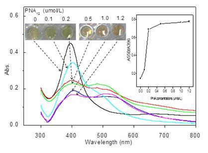 |
| Fig.1: Depicts visual colors of AgNP solutions (0.45 nmol/L) incubated with different PNA12 concentrations (0-1.2 µmol/L) and absorption spectra of corresponding solutions. (© Science China Press)
|
|
A correlated research article was recently published in the journal of Science Bulletin ("A label-free colorimetric assay for detection of c-Myc mRNA based on peptide nucleic acid and silver nanoparticles"), written by Dr. Xia Li from Liaocheng University (Shandong, China), Shandong Taishan Scholar Prof. Jifeng Liu (current, Tianjin Science and Technology University) and Prof. Chenzhong Li at the Florida International University (Miami, USA).
|
|
PNA has the same hydrogen-bonding nucleobases as DNA, but is attached to an uncharged N-(2-aminoethyl) glycine backbone which confers superior target-binding characteristics and physicochemical robustness over native DNA probes. PNA can induce AgNPs aggregations effectively, which reduces the optical absorbance and leads to a significant color change from yellow to red-brown (Fig.1).
|
|
Interestingly, the aggregation of AgNPs can be reversibly controlled by the formation of PND-mRNA complexes by adding c-myc mRNA to the complementary PNS probe-containing solution. The reversible dissociation process could be easily observed by monitoring the color change from red-brown to yellow by the naked eye (Fig.2).
|
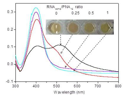 |
| Fig.2: PNA-RNA hybridization disrupts particle aggregation. Photographs of AgNPs solutions recorded after 10 min incubation with PNA12 (100 nmol/L) annealed with its complementary RNA at 0, 25, 50, 100 nmol/L. (© Science China Press)
|
|
The fundamental mechanism of AgNPs-PNA interaction is also investigated at a molecular level. From studying the most probable configurations of PNA structure on AgNPs by molecular modeling, the team found that multiple hydrogen bonds can be formed between PNA on AgNPs, resulting in AgNPs naturally aggregating with one another.
|
|
Additionally, the reduced electrostatic repulsion has also been considered as another factor causing the PNA-induced AgNPs aggregation. In the presence of target c-myc mRNA, the PNA prefers to switch its configuration to combine with the complementary mRNA target, resulting in disaggregation of AgNPs into dispersed Ag nanoparticles.
|
|
"This PNA and AgNPs integrated technology is quite simple, specific and sensitive. We are also considering transferring the liquid-based sensing platform to solid bio-sensing devices such as through a paper or silicon-based platform. In addition, we aim to integrate the bar code technology and smart phone to develop a portable oncogene screening biosensor for point-of-care applications", said Chenzhong Li, professor of Biomedical Engineering at Florida International University, Florida.
|

