| Posted: Jul 11, 2016 |
New focused ion beam strengthens nanotechnology and high-pressure science(Nanowerk News) A new “nano scalpel” enables scientists at DESY to prepare samples or materials with nanometre precision while following the process with a scanning electron microscope. The Focused Ion Beam, or FIB, microscope which has now gone into service also allows a detailed view of the inner structure of materials. The device was purchased by the University of Bayreuth, as part of a joint research project on the DESY campus funded by the Federal Ministry of Research. The FIB will be operated at the DESY NanoLab jointly with the University of Bayreuth. |
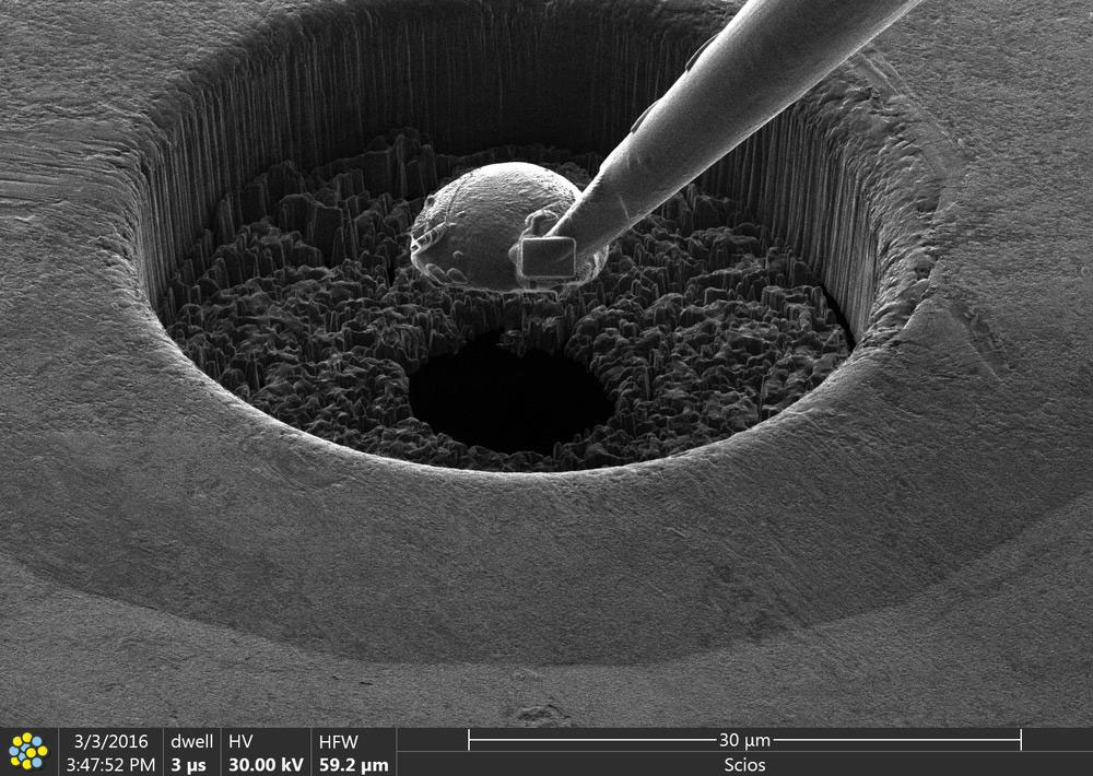 |
| Preparation of a double-staged diamond anvil cell with the focused ion beam microscope. (Image: Leonid Dubrovinsky, Universität Bayreuth) (click on image to enlarge) |
| “The microscope is not only able to examine microscopic defects, cracks or point-like corrosion sites underneath the surfaces of materials, but also to machine the surface of samples with extremely high precision, on a nanometre scale,” explains Maxim Bykov, project scientist from the University of Bayreuth. |
| The ion beam can be used to remove material as though it were a microscopic milling machine; as a result, the combined ion beam and electron microscope is particularly interesting for a wide range of applications in nanotechnology, materials science and biology. |
| “Apart from examining the structure of materials, the ability of the ion beam to remove material also leads to a wide range of different applications,” says Natalia Dubrovinskaia who is a professor at the University of Bayreuth and in charge of the joint research project (No. 05K13WC3). |
| One example is the preparation of tiny diamond anvils, which are used to hold samples during ultra high-pressure experiments. The diamonds used for this are so small that there is no other way of preparing them. The ion beam microscope allows so-called double-staged diamond anvil cells to be prepared with nanometre precision. The ultra high-pressure experiments are carried out at DESY’s Extreme Conditions Beamline (ECB) P02.2, headed by DESY scientist Hanns-Peter Liermann. |
| In addition, the device allows researchers to investigate the chemical composition of samples by measuring fluorescent radiation. |
| “Together with the built-in milling machine, we can not only determine the three-dimensional structure, but also the distribution of the elements beneath the surface by alternately removing material and carrying out a chemical analysis, much like in 3D tomography,” adds Thomas Keller who heads the sub-division microscopy and nano structuring at the DESY NanoLab. |
| Source: DESY |

