| Posted: Oct 07, 2016 |
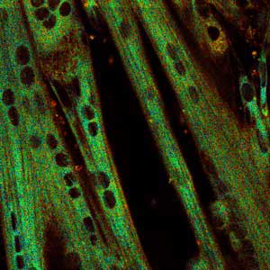
Cellular thermometer(Nanowerk News) A technique that uses fluorescent dyes to measure the temperature inside living cells is helping to reveal the mechanism by which living organisms generate heat ("Direct organelle thermometry with fluorescence lifetime imaging microscopy in single myotubes"). |
| An international team co-led by A*STAR researchers have shown that the dyes, which adjust their light emission in response to temperature, can be used to measure heat generation within muscle cells. The technique has already resolved one controversy over the amount of heat such cells can generate. |
 |
| Fluorescence lifetime imaged on a color scale — at higher temperatures, the fluorescent dye loses its fluorescence more rapidly, which can be detected to measure temperature change. |
| Warm-blooded animals must produce a significant amount of heat to maintain their temperature. Muscle tissue is one source, generating heat through shivering. But muscle cells can also heat up via a chemical process called non-shivering thermogenesis (NST). This process is less well understood, and even the estimates of the amount of heat generated this way by the body can differ wildly. Some tests have suggested they could raise their temperature by one degree Celsius via NST, whereas an estimate calculated from a mass of cells suggested a mere 10-5 degree Celsius per cell. |
| Birgitte Lane and Hideki Itoh at the A*STAR Institute of Medical Biology, along with collaborators in Japan, have unraveled these results using temperature-responsive dyes developed at the Singapore Bioimaging Consortium. |
| The team used ER thermo yellow, a particular dye which sticks to the structure within muscle cells thought to be the site of NST, the sarcoplasmic reticulum. The dye fluoresces after being exposed to light, and the length of time it remains illuminated varies with temperature. |
| Using A*STAR’s analytical fluorescence microscopy facility, the researchers could measure the fluorescent lifetime of the dye with sub-nanosecond accuracy. When they dosed muscle cells with caffeine, they detected a temperature increase of around 1.6 degrees Celsius using ER thermo yellow. When they repeated the experiment using another dye that diffuses throughout the cell, no such temperature increase was observed, confirming the heat was generated at the sarcoplasmic reticulum. |
| “Using this approach allows us to look at heat generation much more accurately within cells, so we can see which cell types have this capability, where in the cell the heat generation is taking place, and then start to dissect the mechanism,” says Lane. |
| The result confirms that specialized cells within muscle tissue are indeed able to raise their temperature by around one degree Celsius via NST. This new methodology could now be used for medical applications such as drug screening, e.g. for obesity, or heat regulation disorders like malignant hypothermia. |
| Source: A*STAR |

