| Posted: Nov 02, 2017 |
Nanosensors demystify brain chemistry
(Nanowerk News) Nanosensors are incredible information-gathering tools for myriad applications, including molecular targets such as the brain. Neurotransmitter molecules govern brain function through chemistry found deep within the brain, so University of California, Berkeley researchers are developing nanosensors to gain a better understanding of exactly how this all plays out.
|
|
During the AVS 64th International Symposium & Exhibition, being held Oct. 29-Nov. 3, 2017, in Tampa, Florida, Markita del Carpio Landry, a professor of chemical and biomolecular engineering, and Abraham Beyene, a doctoral candidate in the Landry lab, will describe their design and use of near-infrared optical nanosensors to image the neurotransmitter dopamine within the brain.
|
|
“Developing sensors for brain chemistry is an exciting area of research that could transform how we diagnose diseases based on imbalances in brain chemistry, such as depression and anxiety,” Landry said.
|
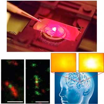 |
| Near-infrared microscopy (top) enables imaging of single-walled carbon nanotube sensors (bottom left) to image dopamine neurotransmission in brain tissue (bottom right). (Image: Markita del Carpio Landry)
|
|
These nanosensors are created by combining carbon nanotubes and biomimetic synthetic polymers with the assistance of sound waves to promote the recognition of a selected small-molecule target. The formed sensors produce a fluorescent signal in the presence of their specified neurotransmitter target. Landry and her team are then able to directly quantify the neurotransmitter levels using the fluorescence intensity as a function of time.
|
|
“These complexes form nanosensors that fluoresce only within the presence of dopamine, a key neurotransmitter implicated in psychiatric disorders and neurodegenerative diseases such as Parkinson’s and Alzheimer’s disease,” Landry said. “We then build microscopes to detect the fluorescent response of the nanosensor so that we can image the nanosensors in living brain tissue.”
|
|
The researchers are already using their sensors to explore how brain chemistry reacts to antidepressants. “We’re seeing some interesting results of how the antidepressant drug Merital affects the way the brain handles dopamine-based neurotransmission,” Landry said. “These key insights may help us to understand how antipsychotics and antidepressants work, and their side effects as well.”
|
|
A simple method to assess brain chemistry is highly desirable for both research and clinical applications. While diseases such as cancer or diabetes are often diagnosed via methods such as blood tests that provide quantitative measurements of imbalances in blood or tissue chemistry, it’s impractical to take a “brain sample” to assess brain chemistry.
|
|
“My lab’s research focuses on the very challenging task of imaging brain chemistry with nanosensors that can report on neurotransmitter concentrations from within the brain and transmit their signals through brain tissue and the cranium,” Landry said.
|
|
Landry and her colleagues are now building a new microscope, a “double infrared excitation-emission” form of fluorescence microscopy for deep brain neurotransmitter imaging, to allow them to image dopamine neurotransmission within the brains of awake and active animals.
|
|
“This will provide us with the capability to determine how antidepressants are affecting brain chemistry and to validate their effectiveness in a living animal model,” Landry said.
|
|
Presentation BI+AS-ThA1, “Engineering and Imaging Excitons for Brain Imaging of Modulatory Neurotransmitters” by Markita Landry, is at 2:20 p.m. EDT, Nov. 2, 2017, in Room 12 in the Tampa Convention Center, Tampa, Florida.
|

