| Posted: Dec 13, 2017 |
New ultra-thin diamond membrane Is a radiobiologist's best friend(Nanowerk News) Depending on the dose and the target, radiation can cause incredible damage to healthy cells or it can be used to treat cancer and other diseases. To understand how cells respond to different doses of radiation, scientists need to direct precise amounts of energy to specific areas of the cell. Measuring dosage can be challenging, however, especially when working with low-energy protons. |
| A collaboration of researchers from the Université de Bordeaux, Centre National de la Recherche Scientifique and CEA-LIST has developed an ultra-thin diamond membrane that can measure the number of protons in a dose of radiation with almost perfect accuracy. The detector attaches to a charged-particle microbeam and enables the delivery of radiation to an area less than 2 micrometers wide. The study, published this week in Applied Physics Letters, ("Cell micro-irradiation with MeV protons counted by an ultra-thin diamond membrane"), represents a valuable technological advance for radiation biology. |
 |
| The ultra-thin diamond membrane detects individual protons as they pass through, allowing researchers to irradiate micron-sized areas on living cells for radiobiology experiments. (Image: Philippe Barberet) |
| Previous experiments had already established that diamond membranes can detect and quantify protons, but until the current study, no one had developed the technology for biological investigations. |
| “The device is completely compatible with living cells in their liquid environment,” said Philippe Barberet, a biophysicist at the Université de Bordeaux. “It will allow us to irradiate different kinds of cells and organisms using single protons, which is not so easy to do using low-energy accelerators.” |
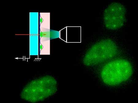
| To test the effectiveness of the diamond membranes when irradiating live cells, the group used a cell line engineered to express a DNA repair protein called XRCC1, tagged with green fluorescent protein (GFP). When DNA damage occurs in these cells, the GFP lights up at the site of the repairs. |
| “XRCC1 is involved in DNA repair pathways and it’s one of the first proteins recruited,” said Barberet. “You irradiate and you immediately see an effect.” They delivered 100 protons spaced 5 micrometers apart to the cells. The resulting pattern of green irradiation spots confirmed that the beam inflicted damage in circles measuring less than 2 microns across. |
| The diamond membranes could become a valuable tool for increasing precision in radiation biology research. The researchers note, however, that their utility is limited to groups who have access to proton beams from particle accelerators. |
| Source: American Institute of Physics |

