| Posted: Dec 15, 2017 |
3D nanoscale imaging made possible(Nanowerk News) Imaging at the nanoscale is important to a plethora of modern applications in materials science, physics, biology, medicine and other fields. Limitations of current techniques are, e.g. their resolution, imaging speed or the inability to look behind opaque objects with arbitrary shapes. |
| However, imaging like this would be useful e.g. for investigating spongy electrodes, thus helping to increase capacity and charging speed of next generation batteries. |
| In a research article published in Nanophotonics ("3D Nano-scale Imaging by Plasmonic Brownian Microscopy"), the team around Prof. Xiang Zhang from the University of California in Berkeley demonstrate a method with stunning properties. |
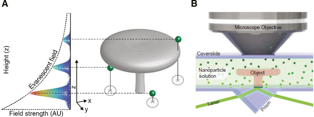 |
| Principle of the PBM technique. (A) PBM relies on three-dimensional localization of freely diffusing nanoparticles (NPs) based on the correlation between evanescent field intensity and height. An object of arbitrary shape (gray) is placed in a solution of randomly diffusing nanoparticles (green spheres), which are illuminated by an evanescent field established by total internal reflection (TIR) at the interface between the substrate and the object. A series of raw images of the particle’s scattering are acquired and the NPs are individually localized in three dimensions as they probe the volume around the object by Brownian diffusion. Images taken at different times record different NPs randomly distributed around the object. The lateral localization is obtained by 2D Gaussian fitting, while the height of particle is estimated by measuring the intensity of the scattering signal. Since the evanescent field decays exponentially with the distance from the interface, the NPs close to the interface will interact with a stronger field and appear brighter than ones further away. Space occupied by the object is inaccessible to the NPs, and the excluded volume corresponds to the object. (B) Experimental setup. The sample, composed of sapphire cover slip with a PMMA structure, is enclosed in a chamber filled with a low-density solution of 50 nm gold NPs whose index is matched to the sample. The chamber is illuminated with a laser and the NP scattering signal is detected using a 100× oil immersion objective. (© De Gruyter) (click on image to enlarge) |
| “We wanted to overcome limitations of current nano imaging techniques and are excited to have found a way to image complex 3d nanostructures even with intricate internal structures such as cavities,” explains Prof. Zhang. |
| Nanoparticles are immersed in a fluid surrounding the object under investigation. By exploiting their special properties when interacting with light, each of the particles acts as a light source, thereby probing the object from all sides, also behind overhangs and inside any cavities. With a resolution of 30nm in all direction, this new technique offers true 3D imaging at the nanoscale. |
| Besides applications in the technology sector, Plasmonic Brownian Microscopy may also be used to map out the biological machinery inside single cells, especially those with intricate internal structures. This would help to further our understanding of the basic mechanisms of living organisms and may give rise to new medical solutions. |
| Source: De Gruyter |

