| Posted: Jan 10, 2018 |
Self-healing of solar cells ecplored with photoconductive AFM(Nanowerk News) A team of researchers have disclosed in which location self-healing occurs for the future solar cells through the use of Photoconductive Atomic Force Microscope (Nano Energy, "Topological distribution of reversible and non-reversible degradation in perovskite solar cells"). |
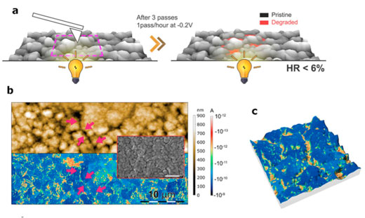 |
| Figure 1: a is the schematic of the microscope used to carry out the experiments. The b is the topography information at the nanoscale, acquired simultaneously with the current map-insert is the SEM image. The c is a 3D rendering composition in which the roughness is the topography information while the colors are related to the amount of photocurrent read. |
| In this work the sample is illuminated from the back of the cell, with the help of a commercially available RGB LED light. While illuminating, a tiny metallic needle is placed on top of the active layer of the solar cell, in which the photocurrent is generated. The needle collects the current which is displayed as a map, blue colors means higher current, and hence a working solar cell. However, the areas with orange colors denote the parts of the cell that are not working. |
| All this information can be gathered with the topography information available from standard topography with the AFM, correlating in which parts of the sample the cell is working correctly. |
| This simple explained methodology is used to disclosed in which parts self-healing occurs. These kinds of cells are formed of little grains, with sub-micrometer scale, which in total, forms the complete solar cell. Each grain is separated from its neighbor, by a “grain boundary”. By using Photoconductive AFM, researchers have proved that grain boundaries acts as seeds in which the degradation starts to spread towards the grain center. |
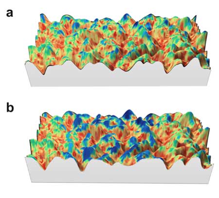 |
| Figure 2: 3D rendering composition in which the roughness corresponds to the topography information while the colors are related to the current information. The a is the degraded sample with a negative applied bias, while in the b a positive bias, just in the center of the image, was applied. It can be seen that on top of the mountains the cell has healed, recovering the level of current when it was new. |
| Even more interesting is the fact that some part of the degradation can be reversed: the cell can be self-healed. This is done by degrading locally the sample using an applied negative bias. Afterwards, researchers apply the contrary bias, in order to recover the initial state of the sample. However, not all the areas are recoverable, only the grains are. Once the grain boundaries are degraded, they do not recover any more. |
| Such fundamental study can have a direct application to device design, as grain boundaries are now spotted as the enemy to avoid the irreversible degradation of the future solar cells. |
| Source: Universitat Autònoma de Barcelona |

