| Posted: May 28, 2018 |
Deciphering the language of cells with a nanophotonic biosensor
(Nanowerk News) Like humans, cells of the same species each have a distinct "personality." When confronted with an external stimulus like a virus, they each secrete a different quantity of molecules and communicate with each other to a varying degree. Studies have already shown that two cells of the same type may not behave identically when subjected to the same treatment.
|
|
In their quest to learn more, researchers from EPFL, working in collaboration with RMIT University in Australia and the University of Lausanne, have come up with an optofluidic device that has a tiny chamber inside. The chamber is around one one-thousandth the size of a raindrop. A cell is placed in the chamber, and then researchers can observe it in real time without disrupting its environment. The quantity and type of the cell's chemical secretions can be monitored continuously.
|
|
The device has been shown to work for 12 hours straight but could function much longer, offering researchers a powerful and innovative selection tool. The results have been published in Small ("Label-Free Optofluidic Nanobiosensor Enables Real-Time Analysis of Single-Cell Cytokine Secretion").
|
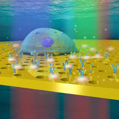 |
| The nanophotonic biosensor developed by the researchers is a glass slide coated with a thin gold film, perforated with billions of nanopores arranged in a precise pattern. (Image: Bo Zhang / EPFL)
|
Studying cells one by one
|
|
All cells function in their own complex way. Cancer cells, for example, produce various hormones and proteins in order to spread and to invade healthy tissue; immune cells respond to an infection or an intruder by secreting chemical mediators called cytokines that stimulate the immune system to fight the enemy. But what is the actual mechanism underlying each cell's behavior?
|
|
Numerous studies have been run on how groups of cells function, but we have precious little information on the behavior of individual cells. Compatible with traditional microscopes, the integrated and miniaturized device developed by the EPFL researchers offers a new way to gain insight into cell processes and communication. It also sets the stage for the development of new therapies to treat cancer and autoimmune diseases.
|
|
"We could, for example, select the most effective immune cells to combat a given disease," says Hatice Altug, a co-author of the study and head of the Bionanophotonic Systems Laboratory at EPFL's School of Engineering.
|
Cells individually housed, fed and analyzed
|
|
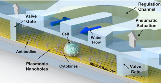
The nanophotonic biosensor developed by the researchers is a glass slide coated with a thin gold film, perforated with billions of nanopores arranged in a precise pattern. A small chamber whose walls are made of porous membranes is placed above the slide. The chamber receives a steady flow of water and nutrients through tiny microfluidic channels. Temperature and humidity are carefully regulated.
|
|
The device contains valves that let scientists insert a cell into the chamber, in which ligands or antibodies are positioned to recognize and capture specific molecules secreted by the cell.
|
|
A broadband light source shines on the chamber. Thanks to an optical phenomenon called plasmons, the nanopores let only one light-wave frequency or color through. When a cell secretes a molecule, it attaches to the antibodies, thereby changing the frequency transmitted by the nanopores. This is how minutes of specific molecules can be identified.
|
 |
| A cell is placed in the chamber, and then researchers can observe it in real time without disrupting its environment. (Image: EPFL)
|
|
The researchers have used their new technique to study cytokine secretion levels in lymphoma cells. "Until now, the methods used to study individual cells have always required fluorophores," says Altug. "Yet these compounds interfere with the cells' functioning and make real-time studies impossible." Maria Soler, the study's co-lead author, adds: "In our device, each nanopore is a separate sensor. Cells can thus settle naturally anywhere in the chamber, and we can analyze them the same way."
|
|
There are numerous potential applications. "Our approach could be used to identify the most aggressive cancer cells in a tumor and decide exactly which treatment to administer to the patient," concludes Xiaokang Li, the study's co-lead author.
|

