| Feb 18, 2019 |
Everything at a glance - new 3D method visualizes nanoparticles in the lung(Nanowerk News) Scientists at the Helmholtz Zentrum München, a partner in the German Center for Lung Research, have presented a new imaging method in the scientific journal ACS Nano ("Three-Dimensional Quantitative Co-Mapping of Pulmonary Morphology and Nanoparticle Distribution with Cellular Resolution in Nondissected Murine Lungs"). |
| This now makes it possible, for the first time, to visualize intact mouse lungs and map the spatial distribution of nanoparticles inside them. The new technique can be used, for example, to measure the efficacy of active substances used in inhaled aerosol therapies. |
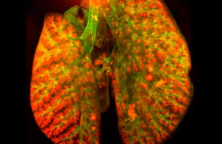 |
| The new technique can be used, to measure the efficacy of active substances used in inhaled aerosol therapies. (Image: Helmholtz Zentrum München) |
| Nanoparticles are tiny particles that can penetrate right through to distant parts of the body. Often, they are associated with adverse health effects, but at the same time novel approaches are also being tested to assess their therapeutic uses, for example as inhaled aerosol drugs. A research team led by Dr. Otmar Schmid, research group leader at the Institute of Lung Biology and Disease at Helmholtz Zentrum München, has now developed a method that will enable a much more thorough examination of the effectiveness of pulmonary aerosol therapy than was previously possible. |
| “In preclinical tests up to now, the lung had to be examined under the microscope in segments – in other words, tissue section by tissue section,” explains study leader Otmar Schmid. “This is very time consuming, doesn’t capture the entire organ and for certain aspects it is not quantitative.” Initially, it was hoped that tissue clearing, a method whereby chemical processes render entire organs transparent, would permit a more detailed examination. Using this technique, the tissue can be illuminated layer by layer and then represented as a 3D image. |
From the trachea to the pulmonary alveoli |
| “Up to now, however, tissue clearing has not been very successful for lungs, as the huge number of alveoli scatter light intensely leading to blurred images”, says Dr. Annette Feuchtinger of the Department of Analytical Pathology at the Helmholtz Zentrum München, who was also involved in the study. “With our new method, it will be possible to circumvent these so-called artifacts. This will enable mapping of the entire airway tree from the trachea to the alveoli without any need for tissue staining. Moreover, we can now correct the tissue clearing dependent distortions which allows us to analyze the 3D structure of the entire lung.” |
| The scientists have already demonstrated the effectiveness of their method in the current study using fluorescent nanoparticles applied to the lung. They were able to visualize their distribution throughout the lung with cellular-level resolution. |
| “Using the new technique, we can precisely measure the dose of nanomedicine delivered to specific – potentially diseased – regions. For instance, this will allow us to optimize the application and the duration of action of nanomedicine and therefore improve potential therapeutic outcomes”, explains Otmar Schmid. “We are thus increasing the knowledge gained from preclinical in vivo models in lung research and, in so doing, improving the development of new drugs for the treatment of lung disease.” |
| Source: Helmholtz Zentrum München |
|
Subscribe to a free copy of one of our daily Nanowerk Newsletter Email Digests with a compilation of all of the day's news. |

