| Dec 02, 2021 |
In the nano-aquarium: Infrared super-resolution microscopy of living cells
(Nanowerk News) Everybody likes gazing at fish in an aquarium and marveling at details such as their colorful skin patterns. In much the same way, physicists at LMU have now developed a sort of nano-aquarium for cells and bacteria (Scientific Reports, "Infrared‐spectroscopic, dynamic near‐field microscopy of living cells and nanoparticles in water").
|
|
Equipped with their novel preparation method and a chemical nanoscope, they managed to visualize patterns in the chemical composition of living cells in their natural aqueous environment.
|
|
The researchers were even able to film the growth and development of an individual cell. This innovation opens up new avenues for the investigation of individual living cells in their environment. “It has the potential to be used in research to fight diseases such as cancer or Alzheimer’s,” says LMU physicist Fritz Keilmann.
|
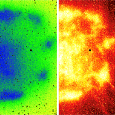 |
| Footprint of a cancer cell (left), infrared image of the same object (right). (Image: LMU)
|
|
The device analyzes the chemical composition along the surface of a cell using local infrared spectra and turns this information into high-precision infrared images. The resolution is far beyond what is allowed by traditional optical microscopy. Directly beneath the metallic needle tip of an atomic force microscope (AFM), a type of scanning probe microscope, the device generates a tiny infrared superfocus, 20 nanometers wide and 40 nanometers deep, and scans over the specimen. To sense their aqueous specimen, the researchers use long-wave infrared light, because only there do all molecules have specific absorption lines.
|
|
“These are therefore called molecular fingerprint spectra,” says Keilmann, member of the Center for NanoScience at LMU. Measured spectra can be automatically assigned to certain molecular groups.
|
|
Keilmann’s team developed the technology, which is already commercially available. The centerpiece of the team’s current research is a sort of super-thin nano-aquarium-window, through which the superfocus of the microscope can shine and map the local chemical composition of cells by means of molecular backscattering.
|
|
Despite its unbelievable thinness of just ten nanometers (only about 30 atomic layers), the nano-aquarium-window is sufficiently robust even at a width of 0.2 millimeters. Even when the researchers dropped living cells on to it from a pipette, it broke neither from the impact nor from the growth and movement of the adherent cells. The thin window deforms locally to a slight extent depending on the local adhesive force of the adherent cell.
|
|
This effect creates a clearly recognizable footprint of the cell in the AFM image, which supplements the infrared image of the chemical composition.
|
|
The Infrared Nanoscopy Group at LMU’s Nanoinstitute (chair of Prof. Stefan Maier) wants to collaborate with biologists in the future to address interdisciplinary questions in the life sciences. Potential subjects could include cell migration and the differentiation of stem cells. It might even be possible to observe aggregation processes of proteins in situ, which play a key role in Alzheimer’s and other neurodegenerative diseases.
|
|
And looking beyond life sciences, the nano-aquarium could also advance our understanding of purely chemical, aqueous processes, such as the corrosion of steel in concrete or the spatio-temporal course of a chemical reaction in close proximity to a nanosized catalyst.
|

