| Apr 29, 2024 |
Microdroplets harness laser light to detect disease markers
(Nanowerk News) A team of researchers led by Nanyang Technological University, Singapore (NTU Singapore) has created tiny droplets that, when activated by laser light, can detect viral protein biomarkers indicating the presence of certain diseases.
|
|
These microdroplets, about one-third the diameter of a strand of human hair, could potentially travel in the bloodstream to reach all parts of the human body and detect particles shed by cells, known as exosomes, which function as disease biomarkers.
|
|
Nanyang Assistant Professor Chen Yu-Cheng from NTU’s School of Electrical and Electronic Engineering, who led the research team with research fellow Dr Fang Guocheng, said the microdroplets could also offer a more precise, effective alternative for photodynamic therapy, which uses light-activated drug carriers to kill abnormal cells.
|
|
The research team’s work was reported in the journal Nano Letters ("Autonomous Microlasers for Profiling Extracellular Vesicles from Cancer Spheroids").
|
Disease detection by hunting for unhealthy cells
|
|
The research team used a liquid crystal to create microdroplets which were then coated with various antibodies that react to different proteins shed by viruses, turning them into disease detectors.
|
|
The microdroplet serves as a focal point for laser light. When the laser enters the droplet, its energy and light are amplified as the laser reflects and bounces inside the droplet repeatedly before exiting the droplet. This creates a stronger energy signal that is emitted from the droplet, leading to more accurate, precise and easily detectable signals.
|
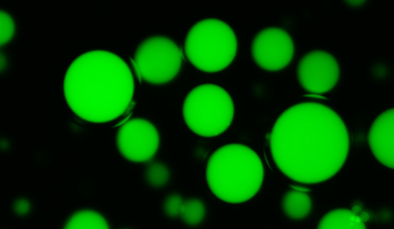 |
| Fluorescence microscopy photo of the laser-activated microdroplets developed by NTU Singapore-led researchers. (Image: NTU Singapore)
|
|
When a microdroplet encounters a protein that reacts with one of its attached antibodies – suggesting the presence of disease or infection – the wavelength of the light reflected out of the microdroplet changes.
|
|
By measuring the wavelength shift as it leaves the microdroplet, researchers have used the technology in lab trials to successfully detect neurological disorders, genetic diseases and cancerous cells.
|
|
Asst Prof Chen said: “Using lasers allows us to amplify subtle biological changes, as they perform well even in scattered or deep tissue environments. Lasers offer strong coherence and intensity and a high signal-to-noise ratio, all of which lead to more precise detection.”
|
|
The researchers said the microdroplets have potential applications in drug screening. “We envision the proposed study can serve as a useful tool for both fundamental biological science and applications such as drug screening and organ or tissue-on-chip applications,” said Asst Prof Chen.
|
|
Currently, tests for diseased cells are done with conventional fluorescent light. Using a laser confers several advantages, said the researchers. The biggest one is greater precision in detecting diseases.
|
|
“As the wavelength of a laser-reflected beam occupies a narrower band than the fluorescence used in conventional tests, the results are clearer and more precise, with less noise and uncertainty,” said Dr Fang, a Presidential Postdoctoral Fellow at NTU’s School of Electrical and Electronic Engineering and the paper’s co-corresponding author.
|
|
“Due to their high sensitivity to changes in the surrounding environment, laser particles have been employed as molecular sensors in various applications,” said Asst Prof Chen.
|
|
These customisable microdroplets also offer flexibility in motion and detection. According to earlier published research, they can be manually controlled using magnetic particles or move autonomously using lipids and surfactants, allowing them to spread within the body. They are also biodegradable and can be safely absorbed by the body.
|
|
“The ability to manipulate microlasers – lasers a few microns in size – in biological fluids opens new possibilities in biophotonic applications,” said Asst Prof Chen.
|
Alternative uses in photodynamic therapy
|
|
The microdroplets could be applied in photodynamic therapy, where patients receive a light-activated drug. These drugs, called photosensitisers, are designed to be absorbed only by diseased or abnormal cells and only take effect when activated by a light source.
|
|
The team’s microdroplets are small enough to navigate the bloodstream and also bind to exosomes. They could be used to deliver these photosensitisers to areas where diseased cells shed exosomes.
|
|
Conventional photodynamic therapy uses an external fluorescent light to activate drug carriers in the bloodstream, which shine light over a large surface area of the body. Doctors can activate the drugs more precisely and locally by using a laser as the light source instead, leading to better targeted efficiency.
|
|
The research team is currently working to develop an integrated biochip which can potentially be commercialised for use in drug screening and bio-assays on a single chip.
|
Additinal Reference
|
|
Paper titled “Motor-like microlasers functioning in biological fluids”, published in Lab on a Chip. DOI: 10.1039/D2LC00513A
|

