| Posted: July 6, 2009 |
An alternative to quantum dots for bioimaging |
| (Nanowerk News) U.S. scientists have developed an in vivo imaging method that offers a potentially safer and more stable alternative to current methods (Upconverting luminescent nanomaterials: application to in vivo bioimaging – free access article). |
| Scott Hilderbrand and co-workers from the Harvard Medical School, Charlestown, have investigated the luminescent properties of yttrium-based nanomaterials and have used the materials to obtain images of blood vessels in mice. |
| The method relies on a process called upconversion, in which particles absorb light of one wavelength and emit light of a shorter wavelength. As Hilderbrand explains, this recent approach to imaging has many advantages over existing methods, such as the use of quantum dots. 'Although very strong emission signals can be obtained, [quantum dots] suffer from potential interference from tissue autofluorescence which can result in poor target to background ratios,' he says. 'Upconversion materials have the potential to eliminate complications from autofluorescence as few, if any, biological components show upconversion luminescence.' |
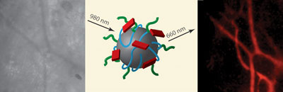 |
| Yttrium oxide nanoparticles give clear upconversion images (right) of blood vessels compared to blue light images (left) |
| Hilderbrand's team used yttrium oxide nanoparticles for the in vivo imaging. The oxide is known to have good stability to light, unlike some imaging materials such as organic dyes. By attaching a polymer coating the team was able to make the particles water-soluble - a requirement for in vivo imaging. The researchers then incorporated a fluorophore on the coating to make the particles luminescent. They found that the particles overcame the problem of autofluorescence and could be used to generate clear images. |
| Manuel Perez, an expert in the field of nanoparticle technologies and molecular imaging, from the University of Central Florida, Orlando, US, says that the work is promising. 'The novelty of the approach is that the nanoparticles are composed of less toxic materials, in contrast to quantum dots,' he explains, suggesting an added benefit over current imaging methods. |
| The researchers suggest that the nanoparticles could find a future use in angiography, intraoperative imaging or other bioimaging applications. |
| Source: Reprinted with permission from Chemical Biology (Ben Merison) |

