| Posted: January 18, 2010 |
Capturing nanometer-scale phenomena with a new optical microscope |
|
(Nanowerk News) Optical microscopes are widely used to observe phenomena on the order of micrometers (µm). In recent years, it has been found that, used ingeniously, an optical microscope can detect phenomena of the nanometer order such as fluorescence generated by a single molecule or light scattered by gold colloidal particles only a few nanometers (nm) in size. In order to understand the mechanisms by which neurons elongate, we have made prototypes of various types of high-precision optical microscopes and observed the motion of the neurons elongating. Our new polarizing microscope [term 1], "Pol-Scope," has enabled the detection of a retardation (a unit for measuring the strength of birefringence) of ca. 0.2 nm and the visualization of the dynamism of actin filament bundles [term 2] with a diameter of 20-60 nm inside a neuron without staining (Fig. 1). Recently, we built a prototype apodized phase contrast microscope of high numerical aperture, and succeeded in direct observations of the actin filament mesh and movements within the cell nucleus. Here, we would like to describe our work, focusing on the apodized phase contrast microscope.
|
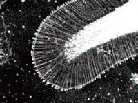 |
| Fig.1 Actin filament bundles at the tip of a neuron (diameter: 20 – 60 nm) as viewed through the new polarizing microscope
|
|
The apodized phase contrast method
|
|
The phase contrast microscope is widely used to observe cells at low magnification. However, this type of microscope suffers from decreased optical resolution due to a halo (a blur of light) around the circumference of a cell that conceals its fine structure. For this reason it is ill-suited to high-magnification, high-resolution observation. The apodized phase contrast method is a phase contrast technique that diminishes the halo by substituting an apodized phase ring (phase ring with a light-reduction film added to its circumference) for the phase ring within the objective lens (Fig. 2). The light-reduction film reduces the low-spatial-frequency component (corresponding to the information for large-size objects), thereby enhancing the high-frequency component (corresponding to the information for small-size objects) in relative terms (Fig. 3).
|
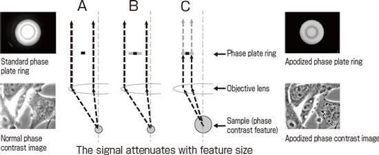 |
| Fig. 2. The principle of apodized phase contrast microscopy (left: phase contrast microscopy, right: apodized phase contrast microscopy)
|
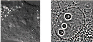 |
| Fig.3 Images of a cell nucleus (left: phase contrast micrograph, right: apodized phase contrast micrograph). Apodized phase contrast microscopy can discern the structure of intracellular particles.
|
|
Application to biological samples
|
|
In 2000, Nikon Corporation developed an apodized phase contrast microscope with halo reduction and applied it to dry objective lenses for low magnification use. We thought that this method would be useful for immersion objective lenses with high aperture and high magnification, and launched a collaboration. Our resulting prototype apodized phase contrast immersion objective lens (NA1.3, 100-power) enabled the visualization of a mesh of actin filaments without staining, a task that is very difficult for a conventional phase contrast microscope (this achievement received the Optics Design "Merit" Prize of the Optical Society of Japan).
|
|
Furthermore, we have prototyped a pupil projection apodized phase contrast microscope, achieving phase contrast viewing with the world's highest numerical aperture (NA1.49). Using this microscope we have succeeded at the direct observation of fine structure inside the cell nucleus, fine structure that a conventional microscope cannot see. The thickness of the optical section was a few hundred nanometers. Going forward we plan to apply this technology for viewing samples from such fields as developmental engineering, medical science, as well as materials science. This microscope allows the detection of a shift in the wave front of light of less than 1 nm, 1/500~1/1000 of the wavelength of the illuminating light (546 nm). We are now looking to develop optical microscopes approaching the resolution of a low-magnification electron microscope.
|
|
Terminology
|
|
Term 1: Polarizing microscope: A type of optical microscope. Illuminating a sample with polarized light enables observation of bifringence. Intracellular microcrystals are detected with high sensitivity, and so it has led to important discoveries such as proof for the existence of mitotic spindles, analysis of DNA structures, and the sliding filament theory of muscle contraction.
|
|
Term 2: Actin filament bundles: Actin filaments, which are formed by polymerization of the protein actin, are an important constituent of the cellular skeleton, and play a variety of roles. The actin fibers form higher-order structures such as bundles and meshes, and play important roles in cell division, extension and differentiation.
|

