| Posted: May 26, 2010 |
Cell imaging: Reporting live on location |
| (Nanowerk News) Visualizing the complex internal cellular processes within our bodies requires the use of photostable, optically active probes that can be tracked with special microscopy instruments. One technique, surface-enhanced Raman scattering (SERS), uses nanoscale tags that maintain emission intensity upon exposure to ultraviolet light—an advantage over conventional fluorescent probes. Now, a team led by Young-Tae Chang from the Singapore Bioimaging Consortium of A*STAR has developed a new approach to generate a library of dyes that can form SERS ‘nanotags’ upon binding with metal nanoparticle colloids ("Combinatorial synthesis of a triphenylmethine library and their application in the development of Surface Enhanced Raman Scattering (SERS) probes"). |
| The SERS technique enhances the vibrational intensity of photoactive dyes known as Raman reporters when they are near gold or silver nanoparticles. Triphenylmethine (TM) dyes, such as crystal violet (CV), have proved useful as Raman reporters under illumination within the visible–near infrared region. Since there is a growing need for additional Raman reporters, Chang and his co-workers undertook a systematic study to synthesize and screen TM dyes using a combinatorial technique commonly applied in drug discovery. |
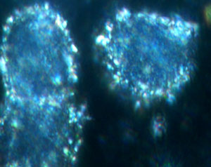 |
| Fig. 1: A dark-field confocal microscopy image of live breast cancer cells with SERS nanotags attached to an antibody. |
| According to Chang, his group was the first to adapt this approach to the synthesis of fluorescent imaging probes. Encouraged by their success, the team applied the same combinatorial technique to other optically active compounds to develop better SERS nanotags. “This is not only about finding better SERS tags; it is also about introducing a new approach to discover them,” says Chang. |
| By immobilizing their starting materials on resin beads, the researchers avoided time-consuming purification steps and toxic reagents such as phosgene gas and chloroform involved in conventional TM synthesis. After linking an initial series of four organic molecules to the beads using a strong base, they reacted the loaded resins with 29 different reagents. Dehydrating the resulting intermediates allowed the researchers to generate the library of TM dyes. |
| After incubating some of these TM dyes with nanometer-size gold colloids to produce nanotags, Chang and his team screened them using confocal microscopy and discovered 13 compounds with higher intensities than CV. They also found that these nanotags tended to aggregate over time in aqueous media, causing significant variations in SERS intensity. To optimize and stabilize the SERS response, they enclosed each nanotag in a polymer shell. This allowed them to identify three biologically relevant nanotags among the shelled nanoparticles. |
| The researchers plan to use their SERS nanotags for in vivo imaging and recently tested them in live breast cancer cells (Fig. 1). They are also developing new nanotags that respond to near-infrared wavelengths for much deeper signal penetration in tissue. |
| Source: A*STAR |

