| Nov 10, 2010 |
Stem cells: Brighten up |
| (Nanowerk News) Stem cells — cells that are capable of differentiating into any type of cell in the body — have attracted widespread interest because of their potential applications in regenerative medicine and developmental biology. In the past decade, scientists have made significant progress in stem-cell research, especially in cultivation and differentiation. Yet despite these advances, there has been little change in the area of stem-cell detection. |
| Immunostaining is currently the most widely used method for detecting stem cells. However, the method is labour-intensive and time-consuming, and involves the use of antibodies that may affect cell physiology. Fluorescent probes — molecules that emit light upon binding specific biological targets — offer an attractive solution to problems commonly associated with immunostaining. |
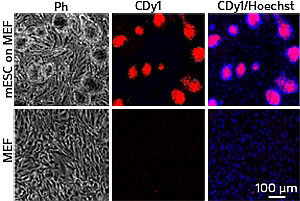 |
| Fig. 1: Microscopy images showing the selective staining of mouse embryonic stem cells (mESCs) grown on mouse embryonic fibroblasts (MEFs) using CDy1. Hoechst staining shows the nuclei of all cells. Only mESCs growing as colonies are stained by CDy1 (Ph, phase contrast image). |
| Young-Tae Chang at the A*STAR Singapore Bioimaging Consortium and co-workers at A*STAR's Genome Institute of Singapore and the National University of Singapore have previously synthesized several diversity-oriented fluorescence libraries (DOFLs) — collections of structurally complex and diverse fluorescent compounds — and applied them to the discovery of novel fluorescent probes for the detection of DNA, RNA and proteins. The team has now screened 280 rosamine compounds in a DOFL and identified a novel fluorescent probe that selectively highlights (stains) stem cells ("A Fluorescent Rosamine Compound Selectively Stains Pluripotent Stem Cells"). |
| The researchers incubated mouse embryonic stem cells and mouse embryonic fibroblasts with each of the different rosamine compounds. They measured the fluorescence of mouse embryonic stem cells and mouse embryonic fibroblasts and selected 20 compounds that displayed the highest differences in fluorescence intensity. Flow cytometry analysis further identified a rosamine compound called CDy1 to have the highest selectivity in staining mouse embryonic stem cells. |
| The researchers were able to discriminate mouse embryonic stem cells from a mixed cell population using CDy1 (Fig. 1). They also found that CDy1 was effective for induced pluripotent stem cells generated from mouse embryonic fibroblasts, making it the first fluorescent probe that can stain both embryonic and induced pluripotent stem cells. Best of all, CDy1 had no effect on the physiology of stem cells, which remained capable of differentiating into other cell types after staining. The findings demonstrate the potential of CDy1 as a novel tool for detecting viable stem cells. |
| "Scientists previously used genetic markers to monitor the production of stem cells, but they could only do this close to the final stage of production. CDy1 allows stem cells to be detected at the early stage of production and therefore provides a means to study the early reprogramming mechanisms of stem cells," says Chang. |
| Source: A*STAR |

