| Feb 03, 2011 |
Boston College receives W.M. Keck Foundation funding for nanoscale optical microscope
|
|
(Nanowerk News) Boston College has been awarded a $1 million grant from the W.M. Keck Foundation to support a team of university researchers developing a new microscope that uses a light-guiding "metamedium" to create images that reveal micro- and macroscopic matter with significantly improved clarity.
|
|
The nanoscale coaxial optical microscope, or NCOM, would join a new class of microscopes known as "superlenses," which function far differently than traditional optical microscopes. These new devices use novel technologies to manipulate light, reconstruct it on computers or assemble bits of images to create one in its entirety.
|
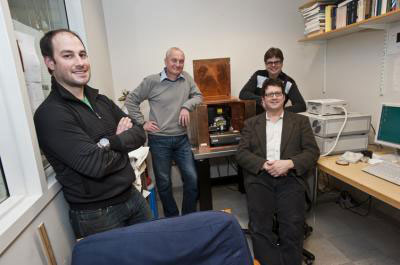 |
| A $1 million grant from the W.M. Keck Foundation will support a Boston College team, including (clockwise from left) Joshua Rosenberg, manager of the University's microscopy imaging facility, Professor of Physics Krzysztof Kempa, Greg McMahon, a researcher and nanolithography specialist in the University's Clean Room Nanofabrication Facility, and Ferris Professor of Physics Michael J. Naughton, the principal investigator. As part of a $1.8 million project, the Keck grant will support the team as it develops a nanoscale coaxial optical microscope, which will use a unique, light guiding "metamedium."
|
|
The NCOM, under development in a $1.8 million project, relies on a bundle of hundreds of nanoscale tubes similar in design to the coaxial cable that supplies TV, Internet and phone signals. The nanocoax design will allow the microscope to focus beams of light on sub-wavelength-sized matter, such as cells or proteins, and then return that light to a camera that presents the image.
|
|
"We're excited by the opportunities this grant from the W.M. Keck Foundation provides and grateful for their support," said principal investigator Michael J. Naughton, Ferris Professor of Physics at BC. "We believe our novel concepts and ideas on microscopy can lead to the development of the nanoscale coaxial optical microscope, which will have a far-reaching impact on scientific investigation."
|
|
Based in Los Angeles, the W. M. Keck Foundation was established in 1954 by the late W. M. Keck, founder of the Superior Oil Company. The Foundation's grant making is focused primarily on pioneering efforts in the areas of medical research, science and engineering and undergraduate education. The Foundation also maintains a Southern California Grant Program that that provides support for the Los Angeles community, with a special emphasis on children and youth. For more information, please visit the foundation website www.wmkeck.org.
|
|
In addition to Naughton, the research team includes Professor of Physics Krzysztof Kempa, Joshua Rosenberg, PhD, manager of the University's microscopy imaging facility, and Greg McMahon, PhD, a researcher and nanolithography specialist in the University's Clean Room Nanofabrication Facility.
|
|
Tiny pieces of matter present a significant challenge to microscopy because they can be smaller than the wavelengths of the electromagnetic waves that strike them. An optical barrier known since the 1800's as the diffraction limit restricts the resolution, or clarity, necessary to distinguishing among the features of matter. Researchers have traditionally used electron microscopes to "see" matter using electron waves, which can be much shorter than light waves, or optical devices after a sample has been colored with fluorescent dyes. Rosenberg says the advantage of the NCOM will be the ability to examine matter using visible light – or photons – without the need to manipulate or stain the sample.
|
|
Where traditional microscopes offer a resolution that allows scientists to distinguish between two features as long as they are separated by 200 nanometers or more, the NCOM is projected to provide resolution of 20 nanometers. Without the need to manipulate or treat the sample, tissue could be examined in its living state, instead of in the vacuum of an electron microscope.
|
|
"With super resolution microscopy, we propose we can achieve 20 nm resolution, which is a 10-fold improvement," said Rosenberg. "That means scientists will be able to see with much finer detail what is going on inside of a cell and how the cell works."
|

