| Feb 09, 2011 |
Novel device sheds light on the beauty of nanoscale science |
| (Nanowerk News) The wonder of science often comes from the endless possibilities opened up by each successive discovery and the unexpected findings that result. Scientists at the University of Bristol now have a new tool that will yield yet more and unprecedented levels of information – and crucially, without disturbing the natural, physical state of the object under scrutiny. |
The past few months have seen physicists at Bristol's Interface Analysis Centre vying for time on the dualbeam instrument, which as centre Director Dr Tom Scott says, "unlocks the key to a whole new world".
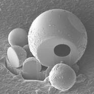 Monmorillonite particles, cut apart to reveal that one of them is hollow. It has so far produced hundreds of images that are as beautiful as they are revelatory, and those at the IAC are keen to see what more the dualbeam can do, working with colleagues from across the University to delve into all matter, from diamonds to insect ears. The dualbeam looks at surface structures with a resolution of less than a nanometre – the equivalent of ten millionths of the thickness of a human hair. The resolution of the images produced is just one nanometre, which is beyond miniscule, given that it takes 1,000 nanometres to make one micron, and 1,000 microns constitute a single millimetre. The dualbeam is so called because it operates using two systems – a focused ion beam (FIB) and a high spec field emission scanning electron microscope (SEM). It operates using gallium ions derived from a liquid metal ion source that are directed at the surface in a tightly controlled beam in which individual atoms are travelling at speeds of up to one million miles an hour. The ion beam can be precisely controlled to remove material from tightly defined areas – essentially performing micro and even nano-surgery on almost any material. |
| Unlike other techniques used for dissecting materials, the dualbeam can extract information and capture images without causing any detectable damage except over a tiny area. It can also deposit materials such as gold and platinum, known for their conductivity, on to the surface structure, providing insights into the composition and behaviour of materials. |
| For physicists looking for quantum wells, biologists looking at the structure of membranes in the ears of tree crickets, and engineers keen to understand the nanostructure of exotic alloys, the dualbeam seems to hold the key to success. |
"It makes things possible which were previously considered impossible, it's at the heart of what makes science beautiful," says Dr Scott. "It can do things in such a precisely defined way to such a high degree of accuracy that it really is incredible. In fact, it's difficult to comprehend just how small a scale this thing works on."
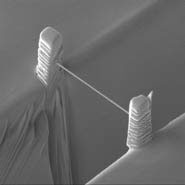 A nano-wire made using ion beam milling for gas sensing applications. It also happens to look like a small-scale version of the Clifton suspension bridge. Some of the project proposals under consideration that would make use of the dualbeam include an examination of the ears of Indian tree crickets, where the dualbeam could be used to slice and view in three dimensions reconstructions of cricket ears. The findings could ultimately inform medical advancements in hearing devices for humans. Another involves examining the materials used to build nuclear power stations. The rate at which they age, and the outputs produced as they do so, is of serious concern. A closer examination of the microstructure of stainless steels, and the processes by which they accommodate strain when affected by thermal cycling in power stations, would yield significant information about potential failure risks that could subsequently be safeguarded against in the design of the next generation of power stations. The dualbeam could also be used in quantum cryptography, to devise ways of transmitting messages in a way that is resistant to attempts to tap into the source, using emitters constructed from a single photonic light source so small and so intricately encoded as to be virtually undetectable. |
| In biochemistry, researchers are looking at making actuators - "gold sandwiches" with a polymer filling which could swim through the bloodstream, collecting information that could be used to inform medical approaches to human disease. |
| Dissecting and reconstructing structures in three dimensions can take a matter of minutes or hours, depending on the volume of the material under scrutiny. The dualbeam also has an automation capability which allows researchers to program it to carry out operational tasks, freeing them up to continue with something else. Dr Scott compares it to a multi-faceted kitchen aid: "This machine basically does all the slicing and dicing, leaving you to concentrate on making a really fantastic meal." |
| Dr Scott is keen to seek out other collaborations that will test the boundaries of every discipline and put materials and this new tool through its paces: "The dualbeam instrument is a clear example of the University's commitment to groundbreaking developments in research. If we are going to be the leaders in the UK and internationally in terms of research we need to be pushing the boundaries of what is technically possible, and this new piece of equipment will certainly enable us to do that." |
| Source: University of Bristol |

