| Feb 21, 2011 |
Atomic structure of nanoparticles measured
|
|
(Nanowerk News) For the first time, scientists have managed to measure the atomic structure of individual nanoparticles ("Three-dimensional atomic imaging of crystalline nanoparticles"). The experimental data could help better understand the properties of nanoparticles in future.
|
|
In chemical terms, nanoparticles have different properties from their "big brothers and sisters": they have a large surface area in relation to their tiny mass and at the same time a small number of atoms. This can produce quantum effects that lead to altered material properties. Ceramics made of nanomaterials can suddenly become bendy, for instance, or a gold nugget is gold-coloured while a nanosliver of it is reddish. Up to now, very little research has been carried out on the effects of these altered properties on living organisms. Only recently, a study caused quite a stir by suggesting that nanoparticles like titanium oxide in toothpaste or sun cream have a similar effect on the human lung as asbestos.
|
 |
|
New method developed
|
|
The exact 3D morphology, atomic structure and especially the surface composition of nanoparticles govern their chemical and physical properties. In a study initiated by the ETH-Zurich scientist Marta D. Rossell from Markus Niederberger's team, professor of the Laboratory of Multifunctional Materials, and the Empa researcher Rolf Erni, the three-dimensional structure of individual nanoparticles has now successfully been determined on the atomic level. The new technique could help improve our understanding of the characteristic of nanoparticles, including their reactivity and toxicity.
|
|
Gentle imaging process
|
|
For their electron-microscopic study, which was published in the journal Nature on 17 February 2011, Rossell and Erni prepared silver nanoparticles in an aluminium matrix. The matrix makes it easier to tilt the nanoparticles under the electron beam in different crystallographic orientations whilst protecting the particles from damage by the electron beam. The basic prerequisite for the study was a special electron microscope that reaches a maximum resolution of less than fifty picometres. By way of comparison: the diameter of an atom measures about one angstrom, i.e. 100 picometres. To protect the sample further, the electron microscope was set up in such a way as to also produce images at an atomic resolution with a lower accelerating voltage, namely eighty kilovolts. Normally, this kind of microscope – of which there are only a few in the world – works at two or three hundred kilovolts. The two scientists used a microscope at the Lawrence Berkeley National Laboratory in California for their experiments. The experimental data was complemented with additional electron-microscopic measurements carried out at Empa.
|
|
Sharpened images
|
|
On the basis of the microscopic images produced, Sandra Van Aert from the University of Antwerp created models that "sharpened" the images and enabled them to be quantified: the images refined by the model made it possible to count the individual silver atoms along different crystallographic directions.
|
|
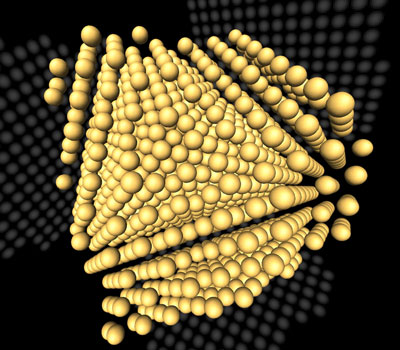
For the three-dimensional reconstruction of the atomic arrangement in the nanoparticle, Rossell and Erni eventually enlisted the help of the tomography specialist Joost Batenburg from Amsterdam, who used the data obtained to tomographically reconstruct the atomic structure of the nanoparticle based on a special mathematical algorithm. Only two images were sufficient to reconstruct the nanoparticle, which consists of around 784 atoms. Finally, two additional experimental projections by Rossell and Erni confirmed the reliability of the reconstruction. "Up until now, only the rough outlines of nanoparticles could be illustrated using many images from different perspectives", says Marta Rossell. Atomic structures, on the other hand, could only be simulated on the computer without an experimental basis.
|
|
"Applications for the method, such as characterising doped nanoparticles, are now on the cards", says Rolf Erni. For instance, the method could one day be used to determine which atom configurations become active on the surface of the nanoparticles if they have a toxic or catalytic effect. Rossell stresses that in principle the study can be applied to any type of nanoparticle. The prerequisite, however, is experimental data like that obtained in the study.
|

