| Mar 10, 2011 |
Researchers use lasers, custom microscope to show gene splicing process in real time
|
|
(Nanowerk News) From neurosurgery to bar code readers, lasers have been used in a myriad of applications since they were first introduced in the late 1950's. Now, with the work being done in Jeff Gelles' Lab at Brandeis University, researchers have developed a way to use lasers to study the splicing of pre-messenger RNA molecules, an essential process in creating proteins to sustain advanced organisms, including human life. This process of splicing is carried out by a cellular micro-machine called the spliceosome.
|
|
"Understanding how these micro-machines function inside the cell is important for many reasons," says Aaron A. Hoskins, a post-doctoral fellow who is a visiting scientist at Brandeis and first author of the paper "Ordered and Dynamic Assembly of Single Spliceosomes," which appears in the March 11, 2011 issue of Science.
|
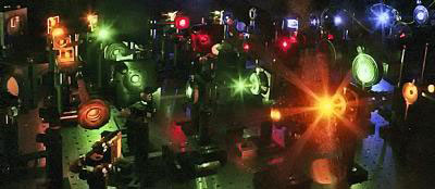 |
| The microscope developed in the Gelles laboratory uses lasers with different wavelengths (colors) to illuminate biomolecules in order to study how cellular micro-machines are assembled. Each fluorescent biomolecule is "turned on" by a different laser, allowing different molecules to be identified and studied simultaneously.
|
|
"One is to further [decipher] basic biology—what makes us humans—and another is to understand how diseases related to these different machines come about," says Hoskins. By understanding how the process works, researchers may eventually be able to come up with therapies that fix the splicing process in cases where it is not working properly.
|
|
The paper reports on a five-year-long collaboration of three research laboratories with diverse expertise to study the splicing process. In addition to Hoskins, authors include: Gelles, the Aron and Imre Tauber professor of biochemistry and molecular pharmacology, whose lab developed the multi-laser imaging system used in the research; Larry Friedman a senior scientist in the biochemistry department who was a key contributor in building the elaborate microscope; Melissa J. Moore, a Howard Hughes Medical Institute Investigator and professor of biochemistry and molecular pharmacology at the University of Massachusetts Medical School and members of Virginia Cornish's laboratory in the Department of Chemistry at Columbia University whose lab synthesized the special dyes that were attached to the spliceosomal proteins so that the proteins could be viewed with the laser microscope.
|
|
"Genomic DNA is sort of like a zip file in that there's a lot of information occupying a very small space," explains Hoskins. "Splicing allows you to decompress the genetic information so the cell can read it before a particular protein is made."
|
|
There are certain regions that code for proteins, called exons, and regions that do not code for proteins, called introns. The regions that do not code for proteins often interrupt the regions that do, therefore they need to be removed—and the remaining pieces must be spliced together—to create appropriate proteins.
|
|
Friedman has spent more than five years developing specialized light microscopes to watch single protein molecules, while Hoskins has been developing the methodology to study these proteins in the complex environments necessary for spliceosome function.
|
|
To view the spliceosome in action -- how it assembles to actually do the splicing—the single yeast components are tagged with florescent dyes then the sample is placed into the microscope. The lasers act as a light source that causes individual tagged molecules to light up so one can actually watch, in unprecedented detail, the splicing process through its various stages.
|
|
"If we have one component of the spliceosome that has a green dye on it and one that has a red dye on it, then we see a green spot and a red spot coming together on the RNA, we know that we are studying part of that assembly process," says Gelles. "By looking at individual molecules one at a time we can actually follow the stages of the assembly process. We can determine whether it happens in the same order on each molecule, or if some spliceosomes assemble differently than others."
|
|
Friedman says that there are easily a hundred or so components that comprise the microscope that he designed and built with his colleagues. There are so many parts, in fact, that it is housed on a platform that looks much like a billiard table, with small tower-like structures and glass lenses scattered throughout.
|
|
"Some pieces were custom made and some are commercial off-the-shelf components that were purchased and put together like an erector set," says Freidman.
|
|
The molecular process known as the "central dogma of molecular biology" concerns the flow of information from DNA to RNA to proteins. RNA contains the chemical information that tells the cells what proteins to make -- for instance, muscle cells use the genes that tell the cell how to make the proteins that are important for muscle, and blood cells use the genes that tell the cell how to make proteins that are important for blood cells.
|
|
With the methods to study the splicesome now at their fingertips, the Gelles lab is also researching the process by which the RNA copy is made, called transcription, and processes by which cells change their shape and move.
|
|
"The thing that's very exciting about this technology is that it's generally applicable to study a wide range of biological problems," says Gelles. "It really enables us to find things out that were very difficult to study using previously existing approaches."
|

