| Aug 29, 2011 |
Amorphous seed nanoparticles show promise to be useful in SERS for magnetic/optical imaging, drug delivery |
| (Nanowerk News) An amorphous-seed mediated strategy has been developed in the Center for Nanoscale Materials Nanophotonics Group for creating bifunctional nanoparticles composed of silver and iron oxide nanodomains ("Plasmonic/Magnetic Bifunctional Nanoparticles"). These hybrid particles exhibit unique optical properties due to surface plasmon resonance from the silver and superparamagnetic responses from the iron oxide. |
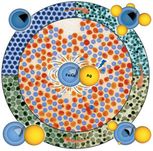 |
| Evolutional pathway from iron particle seeds with thin layers of amorphous iron oxide coating to hybrid nanoparticles composed of solid Ag nanodomains and hollow Fe3O4 nanoshells. Transmission electron microscopy (TEM) images (false colorized) and corresponding schematic illustration (silver: yellow, iron oxide: blue, iron core: black) of the hybrid particles at different stages along the reaction are highlighted on the edge. The TEM image at the center highlights Ag-Fe3O4 hybrid nanoparticles in which Ag and Fe3O4 are false colorized in orange yellow and blue, respectively. TEM analysis was done at Argonne's Electron Microscopy Center. |
| Multicomponent hybrid nanoparticles can exhibit multiple functionalities for applications that are difficult (or even impossible) to achieve from single-component nanoparticles. For example, hybrid noble metal/iron oxide nanoparticles exhibit not only unique optical properties but also magnetic responses. |
| The large-scale synthesis of such hybrid nanoparticles is a challenge. The keys to success for the new amorphous-seed mediated strategy rely on the precise formation of thin amorphous coatings on the seed nanoparticles and strong interfacial adhesion between the two components within each particle. |
| Such multifunctional hybrid nanoparticles are expected to be useful in surface-enhanced Raman scattering (SERS) for chemical and biological sensing, magnetic/optical dual-modal imaging, and drug delivery. Collaborations with scientists in the X-Ray Sciences Division and the Electron Microscopy Center at Argonne National Laboratory as well as the University of Illinois enabled detailed characterization of the materials. |
| Source: Argonne National Laboratory |

