| Posted: February 15, 2008 |
Beautiful nanotechnology images from the SPMage competition |
|
(Nanowerk News) To recognize the continuing contributions that Scanning Probe Microscopes (SPMs) have made to advances in Nanotechnology, an International SPM Image Competition last year identified important and remarkable SPM images.
|
|
An international jury of prominent researchers in the field of SPM judged the images submitted to the Image Prize competition. Here are the top three winners plus some other beautiful entries:
|
|
First place
|
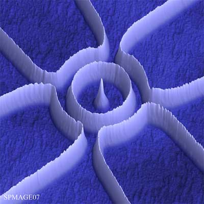 |
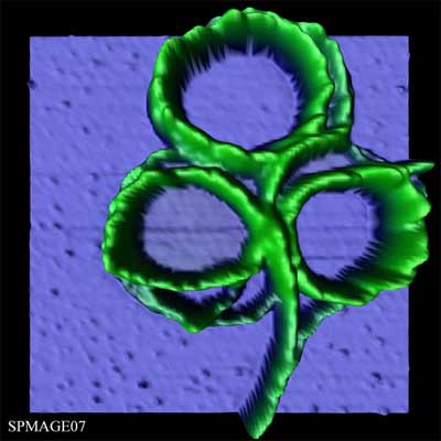
| Nano Rings – The image shows a four-terminal quantum ring structure defined in a two-dimensional electron gas (2DEG) with local anodic oxidation using an atomic force microscope tip. The elevated white lines represent the oxide on the surface of the GaAlAs heterosstructure containing the 2DEG. These oxide lines are on average 15nm high and penetrate just as deep into the sample surface, forming barriers in the electron gas below. The ring has an average diameter of 1 micron and the four outer rectangular areas enclosed by oxide lines are used as in-plane gates to tune the electron density of the four arms of the ring. Measuring Aharonov-Bohm oscillations in the ring conductance this device is used to interferometrically detect the relative phaseshift of Coulomb blockade resonances in two quantum dots induced in the arms of the ring. (Dr Andreas Fuhrer, Nanophysics Group of Prof. Ensslin at ETH Zürich/Switzerland)
|
|
Second place
|
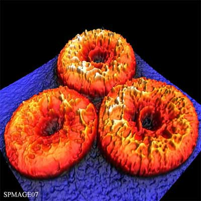 |
| The surface of human red blood cells after treatment with an antibiotic peptide – Phyllomelittin is a novel antibiotic peptide isolated from the skin of the monkey frog Phyllomedusa hypochondrialis. It has been demonstrated that antibiotic peptides exert their activities by disrupting cell membranes. Therefore, the study of the effects of such peptides on cell membranes has been the focus of intense research efforts using the atomic force microscopy (AFM). The aim of this study was to investigate the surface of human red blood cells (RBCs) after treatment with phyllomelittin. The cells were deposited onto a glass slide (blue) and fixed with methanol for 5 minutes. The image shows the intermittent contact mode topography (14.5 µm x 14.5 µm x 819 nm) of three RBCs after 25 minutes of incubation with phyllomelittin at 32 µM. A large number of elevations of few nanometers (yellow) were found to be distributed heterogeneously on the RBCs surface (red), presumably reflecting the regions of the cell membrane disrupted by and/or interacting with phyllomelittin molecules. (Dr Luciano Paulino Silva, EMBRAPA Recursos Genéticos e biotecnología Brasilia/Brazil)
|
|
Third place
|
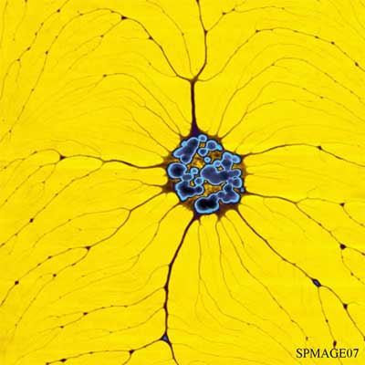 |
| Root – The ability to control the size of conductive nanostructures and to manipulate them on a nanometer scale are priority subjects in the field of nanotechnology. One of the promising way for miniaturization is a template direct method. Polyelectrolytes molecules (PE) offers a range of interesting properties, such as, unique recognition, association and ability to assemble conductive polymers and metals. That is why PE are one of the most attractive templates. The ability to reproducibly create and align well-stretched PE molecules very important for realizing nanoscale electronics. We developed a simple method creating highly aligned PE molecules, which enabled us to straighten and fix PE molecules on the surface without any surface modification or special equipment. Some times, during our AFM measurement, we found unusual structures. One of them have been chosen for SPMAGE07. This image represent part of PE network absorbed on hydrophobic surface. (Mr. Konstantin Demidenok, Leibniz-Institut fur Polymer Forshung Dresden/Germay)
|
|
Other entries
|
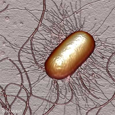 |
| Escherichia coli (E. coli) with Pili and Flagella – An Escherichia coli cell is imaged using tapping mode AFM under dry condition. Well preserved pili and flagella structures can be seen clearly. The size of the cell is about 1.9um long and 1um wide. The width of pili is about 20nm and flagella is about 30nm. (Mr Ang Li, National University of Singapore)
|
|
|
 |
| Nano-Clover – AFM microscopy has emerged as an efficient tool to observe molecules deposited on a surface, specially the changes suffered after induction of external factors. The image shows fibres after treatment with ultrasounds of a bismuth cluster (2 nm high). It is interesting to observe the singular arrangement of the fibres on the surface at the first moments after deposition. (Mrs Lorena Welte Hidalgo, Universidad Autonoma de Madrid/Spain)
|
|
|
 |
| Fantasy – MMX polymers are a particular type of coordination polymer assembled by dimetallic subunits bridged by halides (Cl, Br or I). This kind of compounds is very attractive because of their chemical-physical properties, such as magnetism, electrical conduction, etc… In order to study MMX properties by AFM, the polymers have to be deposited on a surface. The resulting deposition depends on the surface and on the concentration of the sample. In our case, the samples were deposited on a HOPG surface. At low concentrations, the AFM image shows a single MMX polymer, whereas at higher concentrations, superposition of MMXs builds a layer with a peculiar topography, shown in the image. (Dr Rodrigo Gonzalez Prieto, Universidad Autonoma de Madrid/Spain)
|
|
|
 |
| Climatic change on carbon nanotubes – Carbon nanotubes have many characteristics that promise to revolutionize the world of structural materials. There are different ways to grow carbon nanotubes, especially the CVD technique, which allows obtaining SWCNT’s on a silicon surface. These SWCNT can be carried from the silicon surface to another surface, as HOPG, without suffering changes on their properties. That means nanomanipulation of carbon nanotubes. (Mr Miguel Ângel Fernández Vindel, Universidad Autonoma de Madrid/Spain)
|
|
|
 |
| Quantum Forest – GeSi quantum dots on Si, average diameter approx. 70 nm, typical height approx. 15 nm. (Mr Thorsten Dziomba. Physikalisch-Technische Bundesanstalt, Germany)
|
|
To see the entire collection of 51 images go to the SPMage07 site.
|

