| Posted: March 14, 2008 |
Harmonic microscopy |
| (Nanowerk News) Researchers at RIKEN’s Discovery Research Institute in Wako, in collaboration with researchers at Osaka University, have developed a new microscopy method that in principle allows for almost arbitrary enhancements in imaging resolution. |
| The laws of classical optics state that any imaging apparatus can only resolve features that are roughly equal to the wavelength of light used by the microscope. During the past decades, a number of imaging techniques beyond classical optics have been developed that circumvent this limit on resolution. However, many of these techniques achieving ‘super-resolution’ suffer from practical drawbacks—for example, they require close contact with the sample, only allow for planar imaging, or require cumbersome apparatus. |
| The method developed by the research team, led by Osaka University’s Katsumasa Fujita and RIKEN’s Satoshi Kawata, offers a practical way to achieve super-resolution. “The major advantage of our method is that no special skills are required to achieve super-resolution,” comments Kawata. The technique could also be used to take a three-dimensional image from inside a cell. |
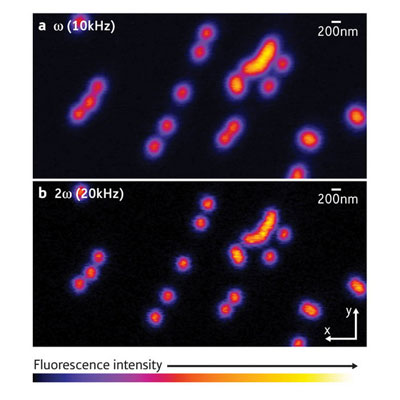 |
| The benefits of higher harmonics. (a) A conventional microscope image of small fluorescent peaks excited with light modulated at a frequency ω of 10 kHz. (b) If the detection frequency is twice the original frequency, a much better resolution is achieved. (Reproduced from Physical Review Letters 99, 228105 (2007), Copyright (2007) with permission from The American Physical Society) |
| Published in the journal Physical Review Letters (High-Resolution Confocal Microscopy by Saturated Excitation of Fluorescence), the new microscopy technique is based on the fluorescence a given sample emits under optical excitation. As the intensity of the optical excitation of the samples in a molecule increases, the fluorescence also increases. At intensities that are too high, however, the fluorescence saturates. Similar to an overexposed photograph, details of the sample structure are lost in the bright emission. |
| Although light that is too bright is detrimental to high-resolution imaging, it also carries components that may be used for super-resolution imaging. If the optical excitation of the specimen is modulated at a certain frequency, the molecules that emit the fluorescence also emit so-called higher-harmonics signals at multiples of the original modulation frequency. The higher harmonics are much weaker in magnitude than the original signal and therefore cannot be saturated as easily, which essentially allows a much better spatial resolution (see images above). |
| As higher and higher harmonics are used, the better the spatial resolution becomes. The only limit to this method is the increasingly low-intensity fluorescence intensity of higher harmonics. However, future developments may bring significant improvements in the detection limit of low signals, and thereby further increase resolution. Indeed, Kawata is convinced that “this practical method will have a big impact on biology and biomolecular sciences, where extremely high imaging resolution in three dimensions is essential.” Efforts are under way to commercialize this technique. |
| Source: Riken |

