| Posted: April 30, 2008 |
Quantum dots illuminate bacterial studies |
| (Nanowerk News) Fluorescent probes are shedding light on bacterial infection. Bradley Smith from the University of Notre Dame, US, and colleagues have made fluorescent probes that can distinguish between different mutants of the same bacterial species and can be used to observe the bacteria in vivo ("Quantum dot probes for bacteria distinguish Escherichia coli mutants and permit in vivo imaging" – free access article). |
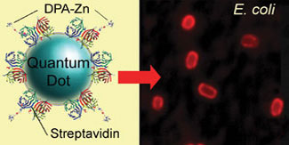 |
| Zinc(II) dipicolylamine coated quantum dots (left) are a selective stain for some bacteria such as a rough strain of Escherichia coli (right) |
| The team made the probes by attaching the bacteria-targeting ligand zinc(II) dipicolylamine (Zn-DPA) to a fluorescent nanoparticle called a quantum dot. Zn-DPA targets bacterial cells because it has a strong affinity for the phospholipids in their outer cell membranes. However, when attached to a relatively large quantum dot, it appears the ligand is unable to reach the phospholipids in some bacteria, leading to selective binding. |
| Smith's team showed that Zn-DPA-quantum dots can stain a rough strain of Escherichia coli intensely, but not smooth E. coli strains or Gram-positive bacteria - a group of bacteria that can be stained by crystal violet dye. They suggest this is because Gram-positive bacteria have thick cell walls with pores too small to allow the quantum dots to pass through. Similarly, the smooth E. coli strains are surrounded by a polysaccharide layer which prevents the Zn-DPA-quantum dots reaching the phospholipids in the membrane beneath. |
| The team says it should be possible to exploit the probes' selectivity in highly sensitive multicoloured staining schemes to identify bacterial species and mutant strains rapidly in contaminated samples. |
| The group also tested the feasibility of using the probes for in vivo imaging of bacterial infection in mice. They found the bacterial fluorescent signal to be 10-fold greater than the background autofluorescence. But, admit the scientists, it is only 1.5 times greater than when bacteria are labelled with Zn-DPA attached to an organic fluorophore rather than a quantum dot. The observed fluorescence is limited because maximum tissue penetration is achieved when a fluorophore's excitation and emission wavelengths are both between 650 and 900nm - these quantum dots emit at 800nm but excite below 500nm. |
| Smith plans to develop the optical imaging method so that it can be used to evaluate antibiotic therapy in animals. 'The challenge is to make very bright and highly selective near-infrared imaging probes that also exhibit favourable pharmacokinetics and low toxicity,' says Smith. |
| Source: Reprinted with permission from Chemical Biology (Freya Mearns) |

