| Posted: May 2, 2008 |
Creating highly sought magnetic nanoparticles in one step |
|
(Nanowerk News) Researchers from the University of Minnesota have demonstrated a one-step technique for producing a class of magnetic nanoparticles that could be used in everything from biomedical applications to data storage. Consisting of an iron and cobalt core with a gold shell, the nanoparticle’s unique, and potentially very useful, magnetic properties were characterized at the NSLS ("FeCo-Au core-shell nanocrystals").
|
|
Magnetic nanoparticles are envisioned for a wide variety of uses. In the medical realm, scientists hope to use single nanoparticles to deliver anticancer drugs or radionuclide atoms to a targeted area of the body, or to enhance the contrast in magnetic resonance imaging (MRI). The particles also could assist in the development of advanced data storage, and further down the road, in spintronic devices. In general, the higher the nanoparticle’s magnetic moment – the measure of a material’s magnetic strength – the more valuable it is for these applications.
|
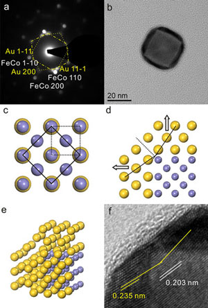 |
| Structure of FeCo-Au nanocrystals. (a) CBED of the nanocrystal in (b), the two distorted hexagons (dot and dot-dash line) indicating two perpendicular Au [011]s. (b) a TEM bright field image of a nanocrystal. (c) illustration of the epitaxial relationship between the bcc FeCo (dotted line) and fcc Au (solid line) (smaller atom, Fe or Cu; larger atom, Au). (d) the perpendicular relationship between the two Au (110)s (indicated by arrows) at two neighboring facets of FeCo {100}, with a twinning at the rim. (e) a 3D illustration of a corner of the FeCo-Au nanocrystal. (f) HRTEM image of a corner of a nanocrystal with the same angle between the Au (111)s from the neighboring Au.. (Image: NSLS)
|
|
“There’s a continuous search for magnetic nanoparticles that perform better than those currently available,” said University of Minnesota Professor Jian-Ping Wang. “Nanoparticles with high magnetic moments are especially valuable for biomedical uses, because the stronger they are, the smaller the quantity that can be used in the body. That’s a very important aspect for clinical trials.”
|
|
Nanoparticles made of iron-cobalt (the core) and gold (shell) – the material recently studied by Wang and his Ph.D. student, Yun-Hao Xu – have a magnetic moment three to four times higher than traditional iron oxide nanoparticles. In addition, the particle’s gold surface is biocompatible and easily links with large molecules – features that make it even more attractive to the biomedical community.
|
|
Although the benefits of these nanoparticles are now well known, Wang and Xu are the first to show how they can be created in a simpler and more efficient manner than traditional techniques. Conventional chemical methods are limited by the solvent’s boiling temperature, which must be extremely high for the reaction to take place, and the risk of exposure to air, which corrodes the sensitive particles.
|
|
“A lot of people like using the chemical method to make nanoparticles from solution,” Wang said. “I prefer the physical method.”
|
|
In their gas-condensation technique, Wang and Xu used a sputter gun to bombard a solid metallic target made of iron, cobalt, and gold with energetic ions. This all happens within a heat bath of high-pressure gas, which transforms the high-energy atoms into the desired nanoparticles.
|
|
The researchers then analyzed the resulting nanocrystals with high-resolution transmission electron microscopy and energy dispersive x-ray spectroscopy, as well as with x-ray magnetic circular dichroism at NSLS beamline U4B. Their findings: the cubic crystals formed through the physical method are very pure, with sharp and clean interfaces between the core and shell; the shell is thick enough to prevent oxidation of the iron-cobalt core (the source of the particle’s unique magnetic properties) for more than four months of air exposure; and the nanoparticles’ magnetic moments are close to the values in bulk iron-cobalt alloy. The details are published in the December 5, 2007 edition of Applied Physics Letters.
|
|
“Our findings suggest that the magnetic nanoparticles formed through this one-step technique are very promising for biomedical applications,” Wang said. “This method can easily be used to synthesize other nanoparticles tailored for uses in different technologies and fields such as for catalysts and hydrogen storage.”
|
|
Currently limited to producing small batches of the particles, the researchers are working on a way to develop the material in more massive quantities.
|
|
Funding for this study was provided by the University of Minnesota’s NanoBiotechnology Initiative and the National Nanotechnology Infrastructure Network, supported by the National Science Foundation.
|

