| Posted: June 19, 2008 |
Nano-Kamasutra and other nanotechnology images |
| (Nanowerk News) The 52nd International Electron Ion and Photon Beam Technology and Nanofabrication (EIPBN) Conference announced six Micrograph Contest winners at its annual Conference held this year in Portland, Oregon May 27-30. |
| The 2008 Grand Prize was won by T. Pinedo and D. Peyrade of the ColloNa team LTM-CNRS in France for a micrograph of self-assembled 1µm and 200nm polystyrene beads taken at 18,000x magnification with an SEM Hitachi 4000 electron microscope. Other winners included micrographs taken with ion microscopes and optical microscopes and a video taken with an electron microscope. |
| "The EIPBN Micrograph Contest attracts many bizarre and beautiful images taken by engineers and scientists in pursuit of cutting-edge technology," said Dr. Randall, vice president of Zyvex Labs. "The most amazing images are often a result of what went wrong and what can be learned from experimentation." |
| Dr. John Randall has been running the contest for the EIPBN Conference for 14 years. Zyvex, a leading nanotechnology company, has hosted the contest since 2001. All of the winners and honorable mentions are available on the Zyvex Labs website at: http://www.zyvexlabs.com/EIPBNuG/uGraph.html. |
| The EIPBN Conference is the world's leading symposium on lithography and nanofabrication. The conference attracts researchers from all over the world to present papers on science and engineering of fabricating, electronic, storage, mechanical, biological, and other devices and structures at the nano-scale. |
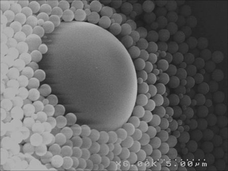 |
| Grand Prize: Begining of Life Description: Self-assembled 1µm and 200nm polystyrene beads. Magnification: (3"x4" image): 18,000X Instrument: SEM HITACHI 4000 Submitted by: T. Pinedo and D. Peyrade. Affiliation: ColloNa team LTM-CNRS. |
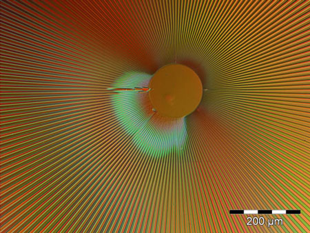 |
| Best Photon Micrograph: Nano Pacifier. Description: Optical micrograph of first imprint at our nanocenter. Pattern is a radial array used for crystallographic orientation for Si wet etch. Image was obtained in polarization mode. Magnification: (3"x4" image): 100 x Instrument: Olympus MX-61 Submitted by: L. Ocola and R. Divan. Affiliation: Center for Nanoscale Materials, Argonne National Lab. |
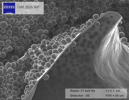 |
| Best Ion Micrograph: Nano Caviar. Description: Spider Eggs on Convoluted Membrane. Magnification: (3"x4" image): 4,000X Instrument: Zeiss ORION Submitted by: John Notte. Affiliation: Carl Zeiss SMT. |
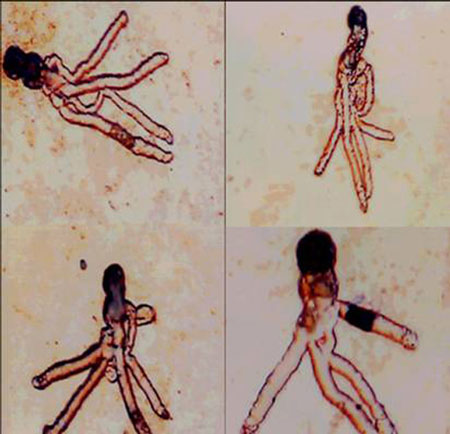 |
| Most Bizarre Micrograph: Kama Sutra. Description: The corrosion protection layer for copper metalization was thick but having pin-holes, so the corrosive stuff entered through the pin-hole (the darkest spot) and then propagated in a branching manner under the inhibition layer (which obviously did not work). Magnification: (3"x4" image): 500x Instrument: Optical microscope Sony AL 100M. Submitted by: Yehiel Gotkis. Affiliation: KLA-Tencor Corp. |
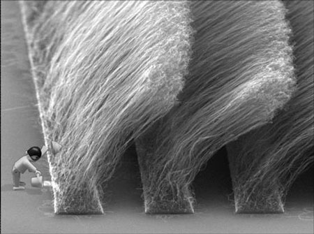 |
| Bending the Rules! Description: CVD grown Carbon nanotubes. Magnification: 12KX Instrument: Philips XL30 Submitted by: Michael Häffner. Affiliation: Universität Tübingen. |
| Source: MRS |

