| Posted: June 23, 2008 |
A look into the nanoscale |
|
(Nanowerk News) Lawrence Livermore National Laboratory researchers have captured time-series snapshots of a solid as it evolves on the ultra-fast timescale.
|
|
Using femtosecond X-ray free electron laser (FEL) pulses, the team, led by Anton Barty, is able to observe condensed phase dynamics such as crack formation, phase separation, rapid fluctuations in the liquid state or in biologically relevant environments. Other Livermore scientists include Michael Bogan, Stafan Hau-Riege, Stefano Marchesini, Matthias Frank, Bruce Woods, former Livermore researcher Sa_a Bajt and former LLNL scientist Henry Chapman, who is now at the Centre for Free Electron Laser Science, DESY, in Hamburg, Germany.
|
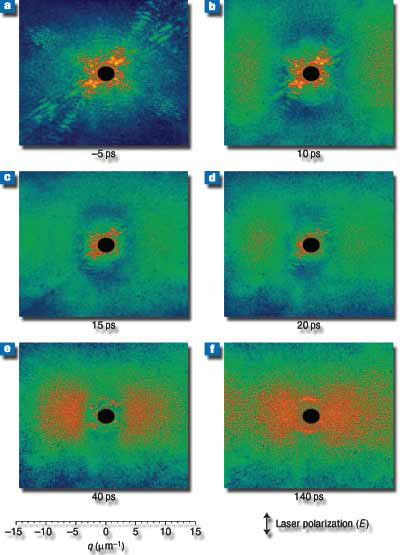 |
| Sample evolution revealed by coherent X-ray diffraction.
Measured single-shot diffraction patterns at 25 ps (a ), corresponding to the object just before the laser excitation pulse, and diffraction patterns from the same object at 10 ps (b), 15 ps (c), 20 ps (d) 40 ps (e) and 140 ps ( f ) after the laser pulse. (Image: Lawrence Livermore National Laboratory)
|
|
"The ability to take images in a single shot is the key to studying non-repetitive behavior mechanisms in a sample," Barty said.
|
|
As the femtosecond laser blasts the sample, it is destroyed, but not before the scientists created images with a 50-nanometer spatial resolution, and a 10-femtosecond shutter speed. (A femtosecond is one billionth of one millionth of a second. For context, a femtosecond is to a second as a second is to about 32 million years.)
|
|
"This experiment opens the door to a new regime of time-resolved experiments in mesoscopic dynamics," Barty said. "This technique could be extended to a few nanometers spatial and a few tens of femtoseconds temporal resolution."
|
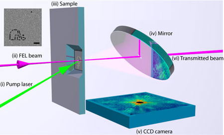 |
| A visible light laser beam (i) is focused onto the sample (iii) and acts as the excitation pulse. A soft X-ray pulse (ii) is focused to the same location but at a continuously variable delay. The X-ray pulse diffracts from the sample, carrying information about the transient sample structure to the CCD detector (v) in the form of a coherent diffraction pattern. A mirror (iv) separates the direct beam from the diffracted light: the direct FEL beam (vi) passes straight through a hole in the mirror and is not detected in the CCD image. (Image: Lawrence Livermore National Laboratory)
|
|
This is the first time that optical pulses have been used to image samples at the nanometer-spatial resolution scale. Earlier studies were limited to a few micrometers.
|
|
The "shutter speed" of the measurements is determined by the femtosecond duration of the FEL X-ray pulse. This allowed the team to obtain nanometer spatial resolution of violent and destructive events in which the sample is completely destroyed.
|
|
The new technique is necessary to study ultrafast dynamics of non crystalline materials at nanometer-length scales. This includes fracture dynamics, shock formation, spallation, ablation and plasma formation under extreme conditions. The technique also allows researchers to image dynamic process in the solid state such as nucleation and phase growth, phase fluctuations and various forms of electronic or magnetic segregation.
|
|
The research appears in the June 22 online edition of Nature Photonics.
|

