| Posted: July 31, 2008 |
X-Ray diffraction of aerogel reveals nanoscale 3-D bulk lattice structure |
|
(Nanowerk News) A multi-institutional team of scientists has used beamline 9.0.1 at the Advanced Light Source to perform high-resolution x]ray diffraction imaging of an aerogel for the first time, revealing its nanoscale three-dimensional bulk lattice structure down to features measured in nanometers.
|
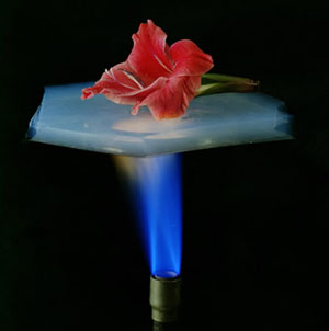 |
| Aerogels, sometimes called "frozen smoke," can be made from different materials. This silicon aerogel is an efficient insulator.
|
|
Aerogels, sometimes called "frozen smoke" or "San Francisco fog," are nanoscale foams: solid materials whose sponge-like structure is riddled by pores as small as nanometers across and whose strength is surprising, given their low density. Many porous materials are extraordinary for their properties as insulators, filters, and catalysts; they are used to produce clean fuels, to insulate windows and even clothing, to study the percolation of oil through rock, as drug-delivery systems, and even to cushion the capture of high-velocity comet fragments in outer space.
|
|
"The smallest pore size is the key to the strength of porous materials and what they can do," says Stefano Marchesini, an ALS scientist at Berkeley Lab, who led the research. "Seeing inside bulk porous materials has never been done before at this resolution, making this one of the first applications of x-ray diffractive microscopy to a real problem."
|
|
Team members from Lawrence Livermore National Laboratory, the University of California at Davis, Arizona State University, Argonne National Laboratory, and Berkeley Lab performed the x]ray diffraction imaging and have published their results online in Physical Review Letters (" Three-Dimensional Coherent X-Ray Diffraction Imaging of a Ceramic Nanofoam: Determination of Structural Deformation Mechanisms").
|
|
Seeing inside foam
|
|
One way to study aerogels and other nanofoams is with electron microscopy, which can image only thin, two-dimensional slices through the porous structure of the material. Another method is straightforward x-ray microscopy, using zone plates as "lenses"; microscopy can penetrate a sample but has difficulty maintaining resolution at different depths in the material. Small-angle x]ray scattering (SAXS) can also gather limited structural information from finely powdered aerogels, but SAXS cannot provide full three-dimensional information. None of these techniques can capture the 3-D internal structure of nanofoam samples measured in micrometers, a few millionths of a meter across.
|
|
X-ray diffraction approaches the problem differently. A laser-like x-ray beam passes all the way through the sample and is diffracted onto a CCD detector screen; diffraction patterns are repeatedly stored while the sample is moved and rotated. A typical series requires approximately 150 views in all.
|
|
The individual diffraction patterns are then processed by a computer. The way the photons in the beam are redirected from each component of the structure is different for each orientation, and comparing their intensities serves to position that component precisely in three-dimensional space. Thousands of iterations are required | in the present study, team member Anton Barty of Livermore led the solution of almost 100 million measured intensities, as opposed to the 100 thousand or so typical of, say, protein crystallography | but the end result is a 3]D image of the tiny sample at nanometer-scale resolution.
|
|
Foam-like structures are described in terms of interconnecting lattice beams and the nodes where they intersect. These elements became vividly apparent in the reconstructed 3-D images of the aerogel used in the imaging at the ALS, which was made of tantalum ethoxide (Ta2O5), a ceramic material proposed for cladding capsules of hydrogen isotopes for inertial-confinement fusion experiments being pursued at Livermore.
|
|
"The strength and stiffness of foam-like structures are expected to scale with their density, relating the density of individual elements like beams and nodes to the overall density," Marchesini says. "But below about 10 percent density, the strength of aerogels like the ones we tested | on the order of 1 percent density | is orders of magnitude less than expected."
|
|
Of the theories that seek to explain this phenomenon, one is the "percolation" model, in which fragments become detached from the load-bearing structure and add mass without contributing to strength. The alternate "heterogeneities" model proposes that the structure is increasingly riddled with defects like micron-sized holes and buckles more easily.
|
|
A third theory is the "diffusion-limited cluster aggregation" model: blobs of material accumulate that are connected by thin links, instead of sturdy beams between nodes.
|
|
"The high resolution we achieved allowed us to see which of these models more accurately described the actual observed structure," Marchesini says. By seeing the foam from the inside, the team was able to see exactly how it was structured, and the shape and dimensions of each component. "The structure was far more complex than anything we'd seen in earlier images obtained using this technique."
|
|
What the team observed in their 3-D images of the tantalum ethoxide aerogel was a "blob-and-beam" structure consistent with the third model, that of diffusion-limited cluster aggregation. The observed structure explained the relative weakness of the low-density material and also suggested that changes in methods of preparing aerogels might improve their strength.
|
|
Into the future
|
|
"We'd like to use x-ray diffraction to study a range of porous materials and nanostructures in general, for example porous polymers developed at the Molecular Foundry for storing hydrogen as fuel | and at even higher resolutions," Marchesini says. "To do so, David Shapiro, who built the end station we used for this work, is working with us to overcome some obstacles."
|
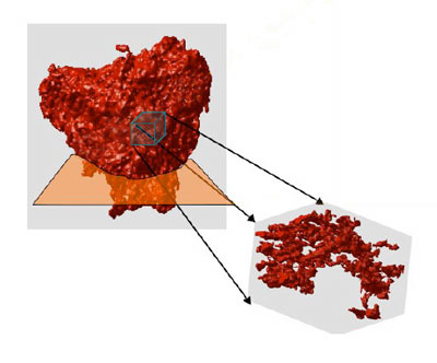 |
| A 500-nanometer cube of aerogel from the interior of the 3-D volume, reconstructed by x-ray diffraction. The foam structure shows globular nodes that are interconnected by thin beam- like struts. Approximately 85 percent of the total mass is associated with the nodes; relatively little of the mass is in the load-bearing links.
|
|
One is time. At present, each sample takes months of work. After preparation, the experiment first requires one or two days of mounting, rotating, and exposing the sample to the x-ray beam, about a minute per view | because of a slow detector | for 150 views. There follow weeks of computation time. "And after all this, you can find out the sample was no good, so you have to start over," Marchesini says.
|
|
Improved sample handling, faster detectors, and a beamline dedicated to x-ray diffraction are principal goals. The Coherent Scattering and Diffraction Microscopy (COSMIC) facility, a top priority in the ALS strategic plan, will provide intense coherent x-rays with full polarization control.
|
|
"We are also collaborating with Berkeley Lab's Computational Research Division to develop efficient and robust algorithms to speed up the time needed to construct the 3-D image from the individual rotated views," Marchesini says. "This will open an entire spectrum of possibilities for new ways of seeing the very small | not just aerogels but virtually any unknown object, from nanostructures to biological cells."
|
|
This work was principally supported by the Department of Energy through a variety of grants, by Laboratory Directed Research and Development programs at Livermore, and additionally by the National Science Foundation.
|
|
Additional information
|
|
"Three-dimensional coherent X-ray diffraction imaging of a ceramic nanofoam: determination of structural deformation mechanisms," by A. Barty, S. Marchesini, H. N. Chapman, C. Cui, M. R. Howells, D. A. Shapiro, A. M. Minor, J. C. H. Spence, U. Weierstall, J. Havsky, A. Noy, S. P. Hau-Riege, A. B. Artyukhin, T. Baumann, T. Willey, J. Stolken, T. van Buuren, and J. H. Kinney, appears in Physical Review Letters online publication and is available to subscribers at http://link.aps.org/abstract/PRL/v101/e055501.
|

