| Posted: August 5, 2008 |
How to clean a Transmission Electron Microscope without disassembling it |
|
(Nanowerk News)
|
|
Shin Horiuchi, the Research Group for Nanoscientific Measurements, the Nanotechnology Research Institute of the National Institute of Advanced Industrial Science and Technology (AIST) and Consult Zero Loss Imaging Co., Ltd. (CZI) have jointly developed a process for cleaning a transmission electron microscope (TEM), and for preventing samples from being contaminated during the electron beam irradiation.
|
|
Though transmission electron microscopes have been widely used to investigate structures at the nanometer level in the various fields such as materials and biotechnology, contamination of specimens induced by electron beam irradiation has been a problem to impair the quality and reliability of analyses. This problem is mainly caused by hydrocarbon-based contaminants remaining in the microscope after prolonged use. The Japanese team has succeeded in removing such contaminants without disassembling the microscope by circulating activated oxygen radicals inside the microscope generated from the plasma generator.
|
|
As a result, specimen contamination caused by the electron beam irradiation is decreased and the performance of the analysis is improved. Because the contaminants are hydrocarbon-based, the cleaning is especially effective when analyzing light elements in organic materials such as polymers. The technique is also effective for analysis techniques requiring long time beam irradiation such as electron tomography, nanobeam electron diffraction, EELS (electron energy-loss spectroscopy), EDX (energy dispersive X-ray diffraction) and STEM (scanning transmission electron microscope), and thus it will contribute to advanced analysis techniques.
|
|
Details of the research will be announced at Microscopy & Microanalysis 2008 which will be held on August 3-7, 2008 by the American Microscope Society in Albuquerque, U.S.A.
|
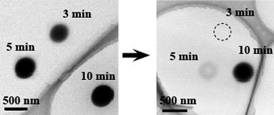 |
| When the electron beam was irradiated on a carbon thin foil, which was used as a test specimen, a dark spot was formed on the irradiated region of the specimen. This contamination spot was produced by 3 min irradiation before the cleaning of the TEM column (left side). After cleaning, the contamination was barely seen even for the 5 min irradiation. Although the 10 min irradiation seems to produce heavy contamination, they were ten times thinner than the contamination before the cleaning (right side). (Images: AIST)
|
|
Background for Research
|
|
Although the recent developed TEMs have realized atomic scale resolution, they cannot frequently perform the full abilities especially when analyzing polymers and organic materials at the nanometer level. Many of the problems do not come from the electron microscopes themselves, but from the specimen contaminations. The fact that the observation performance (effective resolution) and chemical analysis performance (minimum detection limit) are reduced by the contamination (dirt, non-objective substances) deposited on the specimen as dark dots in the irradiated region with several 10 nm diameter.
|
|
Many molecules are floating inside a transmission electron microscope, even though it is maintained at high vacuum. The oil of the vacuum pump, leakage of the vacuum, evaporated molecules originated from the degradation of specimens are accumulated on the inner walls of the microscope over many years of use. Some of these accumulated contaminants become gaseous and floats in the vacuum again. Hydrocarbon compounds were mainly included in these molecules. When the electron beam is irradiated, the hydrocarbon molecules are excited and deposited on the specimen being observed as contamination. Although it has long been desired to clean the inside of electron microscopes to reduce such contaminations, the only possible way to date is to clean the each parts of the microscope by disassembling.
|
|
History of Research
|
|
AIST has been employing transmission electron microscopy to analyze the local structure of organic materials such as polymers at the nanometer level. Through the research, it has been found to be important to control specimen contamination at the molecular level in order to analyze the chemical state of organic materials.
|
|
To solve this problem, AIST collaborated (FY2007-2008) with CZI to develop a non-destructive system of cleaning transmission electron microscopes, as part of the Industrial Technologies Research and Development Project (small and medium-sized business support) commissioned by the Ministry of Economy, Trade and Industry. The scientists aimed to develop a cleaning technique using chemical etchings by activated oxygen radicals. In addition, by using the technique they also aimed to maintain and improve the performance of observation and analysis using the transmission electron microscope without disassembling at low cost.
|
|
Details of Research
|
|
The contaminants formed when the electron beam is irradiated to a test sample (carbon thin film) in a transmission electron microscope used for five years is shown in the figure on the left. A clear black dot was formed after electron beam irradiation for three minutes, and the contaminant became thicker with longer exposure time such as five minutes and ten minutes. Thus, when the contaminant attaches to the specimen, high-resolution observation and chemical analysis become difficult because the thickness increases locally by the electron beam irradiation. Especially, when many organic samples are observed, the microscope becomes severely contaminated due to damage of the specimen caused by electron beam irradiation. As the main element of the contaminant is carbon, performance of analyzing organic specimens, which contain mainly carbon atoms, is deteriorated.
|
|
To remove such contaminations in the microscope without disassembling it, the team developed a technique for circulating activated oxygen radicals within the microscope. This chemically decomposes contaminants and removes them to the outside. Because the vacuum passage of a transmission electron microscope is extremely narrow compared with other vacuum devices, it was difficult to obtain a sufficient flow of the short-lived activated oxygen radicals while maintaining a moderate level of vacuum necessary for plasma generation. Through trial and error, the researchers obtained a large flow quantity of active oxygen and stable generation of plasma.
|
|
The Japanese team optimized the conditions of pressure and time to produce the active oxygen flow necessary for cleaning the microscope and thus established an effective cleaning system. When the electron beam is irradiated to a sample in the same condition after cleaning the inside of the microscope by operating the system automatically for two hours, the black dot was reduced dramatically. No black dot was generated at all after irradiation for three minutes as shown in the figure on the right above, and was generated only slightly after irradiation for five minutes. The thickness was 1/5 compared with that before cleaning, though a clear black dot was confirmed after ten minutes.
|
|
The scientists aim to construct a system that the user can properly use according to the purpose and situation, as the frequency of cleaning depends on the purpose and the type of specimens. The cleaning greatly improves the reliability of the analysis data and the accuracy of images of organic samples such as polymeric materials, etc. at the molecular level. It was confirmed that there was no deterioration the electron microscope such as O-rings for vacuum, even though the decomposition of organic substances by active oxygen was used. The researchers also consider that the exhaust gas has no environmental impact because the life of activated oxygen radicals is about 1/100,000 second.
|
|
Future Schedule
|
|
Our newly developed technique for cleaning transmission electron microscopes can maintain a clean state in which the sample cannot easily be contaminated within the microscope. The technique is expected to be effective for structural analysis of polymeric materials such as rubber and plastic at the molecular level. The scientists will prove the effectiveness of this technique by analyzing various samples in cooperation with companies and universities.
|

