| Posted: August 18, 2006 |
More details in the nanocosmos of the cell |
|
(Nanowerk News) Scientists at the Max Planck Institute for Biophysical Chemistry have further opened the door to the nanocosmos of the cell: The researchers have, for the first time, improved the resolution of STED microscopes (Stimulated Emission Depletion) to 15 nanometers. Already in April, they achieved a detail sharpness of 60nm in cells with the microscopes whose principle was developed by them only a few years ago. The scientists can now image more details than before from the inside of a cell as they have further decreased the effective focal spot of the STED microscope. The required intensive light for this purpose can be used only because a new trick prevents the fluorescent dyes from extensive bleaching. ("Macromolecular-scale resolution in biological fluorescence microscopy")
|
|
How viruses infect a cell, how nerve cells transport signals or how proteins work - the nanocosmos of nature remains hidden to the human eye. However, in order to still be able to perceive the seemingly invisible, we need to enlarge the object - for example, with a fluorescence microscope. Fluorescent markers are attached to proteins and other biomolecules so that scientists can observe the marker. For a long time, low resolution prevented a deeper look into the function of proteins - single proteins with their dimension of 2-20 nanometers diameter were, until now, just too small.
|
 |
|
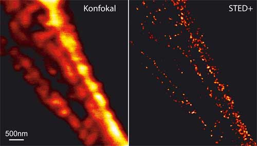
A look at the inside of cells becomes sharper: Both figures above show the filaments in a human nerve cell; left with a common confocal microscope, right with a STED microscope plus mathematical deconvolution. The resolution of the STED microscope is better by more than an order of magnitude. (Image: MPI für biophysikalische Chemie)
|
|
Scientists at the Max Planck Institute for Biophysical Chemistry in Göttingen have now achieved a resolution of up to 15nm with their STED microscope (Stimulated Emission Depletion). Their fluorescence microscope is thereby twelve times sharper than a conventional one. Already in April, the team of scientists lead by Professor Stefan Hell achieved a detail sharpness of up to 60 nanometers in cells.
|
|
Only a few years ago, physicists believed that it was impossible to resolve details that lie closer together than 200 nanometers . This limit is imposed by Abbe’s Law, whereby the resolution of a light microscope cannot be more accurate than half of the wavelength of light entering the microscope.
|
|
Hell and his co-workers have overcome this limit with a trick. They excite the fluorescence dyes that have been attached to the proteins with a blue light beam. The size of the spot from this beam though cannot be made sharper than 200 nanometers, as governed by Abbe’s Law. However, before the excited molecules in the light spot can fluoresce, the molecules in the outer section of the light spot are forced to relax. To this effect, they overlap a second ring-shaped light beam, the STED beam, over the excitation spot. This now means that only those molecules in the clearly smaller spot in the center of the light ring remain excited and can therefore finally glow.
|
|
The more intense the STED beam, the smaller is the spot in which the molecules can still fluoresce. The resolution is also therefore better. However, the fluorescing dye molecules also bleach faster in an intense light beam and one sees - nothing. The Göttingen-based scientists discovered that the fluorescing molecules bleach for the most part because they are continually transferred for a microsecond into a further dark state: physicists speak of a triplet state. For a molecule in this state, when hit by a light particle, it would be irreversibly bleached. The solution to the problem is to irradiate the molecule with a light pulse that allows 4 microseconds between the pulses - enough time for the molecules to return from the dark state. Subsequently, the molecules are available once again for excitation and relaxation.
|
|
"The full potential of the STED technique has still not been fully exploited," according to Professor Hell. Resolution of the size order of a dye molecule is imaginable - this corresponds to a sharpness of one or two nanometers. Fluorescence microscopy is most often used in biology. Its advantage lies in the fact that the inside of living cells can be observed without destroying the cell. With their STED technique, the scientists in Göttingen have already shown how the protein Bruchpilot is spatially concentrated in synapses, and how it triggers the building of active synaptic zones at which the nerve cell selectively releases neurotransmitter. Furthermore, they explored how, during synapse, the released proteins assemble at the presynaptic membrane.
|

