| Nov 25, 2012 |
Analysing spider venom |
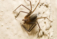
| (Nanowerk News) A European project investigated the effect of brown spider venom on the structure and biophysical properties of cellular membranes. By using state-of-the-art fluorescent techniques, scientists succeeded in directly visualising venom-induced changes in cells. |
 Biting from the brown spider of the Loxosceles genus causes several clinical manifestations in mammals, especially necrotic skin degeneration, haematological disturbances and renal failure. The venom of the Loxosceles spiders combines a toxic component with a rare enzymatic activity, termed sphingomyelinase D (SMD). SMD catalyses the conversion of the cell membrane constituent sphingomyelin (SM) into ceramide-1-phosphate (Cer-1-P), which is implicated in various signalling pathways. While SM is found in abundance in the vascular epithelium and in red blood cells, the concentrations of the product Cer-1-P are very low. At present, the precise mechanism of venom action is incompletely understood. Seeking to delineate this, the EU-funded project ‘The study of membrane phenomena caused by sphingomyelinase D from spider venoms’ (Enzymembrane) worked on the effect of SMD on cellular membranes. More specifically, project scientists investigated the structure and dynamics of model membranes containing SM and Cer-1-P by using artificial lipid bilayer membranes in the shape of giant unilamellar vesicles (GUVs). Laser scanning confocal fluorescence microscopy (LSCFM) was employed to examine biochemical and biophysical aspects of enzyme action by directly visualising fluorescently labelled SM molecules. Analysis of the model GUV membranes containing SM showed alterations in cell membrane morphology associated with SMD action. Red blood cell membranes showed additional fusion events. |
| Collectively, the Enzymembrane project results generated new information on the activity of spider venom on cell membranes, unveiling the mechanism of spider venom toxicity. The combinatorial approach could be utilised to explore enzyme-membrane interactions in other cellular systems. |
| Source: Cordis |

