| Jan 24, 2018 |
Vitamin C in the body can be tracked by fluorescence
|
|
(Nanowerk News) Vitamin C is best known as a nutrient. In high enough doses, however, vitamin C also shows potential against many cancers, according to recent studies. To successfully develop vitamin C (chemically named ascorbic acid) as a medication, it is crucial to probe its concentration in the body, thus ensuring safe and effective doses.
|
|
Monitoring the changing levels of a delivered drug is not easy. For ascorbic acid, one method is to use the reaction between the compound and fluorescent probe molecules, allowing chemists to visually trace its movement through the body. Fluorescence requires light, however, and existing probes use light of the wrong wavelength to penetrate living tissue. Now, a team led by The University of Tokyo's Institute of Industrial Science (IIS) has developed a new probe that could shine a path to ascorbic acid as a cancer treatment.
|
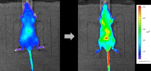 |
| The mice were injected with a dose of extra Vitamin C, raising the concentration above normal. The probe signal spread throughout the body over one hour, becoming particularly intense in vital organs. This is the first time that researchers have directly imaged Vitamin C administered to a mouse. (Image: Kazuyuki Ishii, Institute of Industrial Science, The University of Tokyo)
|
|
In a study reported in Scientific Reports ("In vivo fluorescence bioimaging of ascorbic acid in mice: Development of an efficient probe consisting of phthalocyanine, TEMPO, and albumin"), ascorbic acid levels in mice were tracked using a complex molecule called R2c. Using some clever chemistry, the IIS team designed this probe to react with ascorbic acid molecules in the body. An hour after R2c was injected into mice's tails, the entire body was fluorescent, highlighting the ascorbic acid naturally present in the mice, as the R2c probe was circulating through the bloodstream.
|
|
Next, the mice were injected with a dose of extra ascorbic acid, raising the concentration above normal. Within 10 minutes, the fluorescence signal flared up in the abdomen - showing that this was the first destination of the ascorbic acid dose flowing from the tail. Like before, the probe signal then spread throughout the body over the next hour, becoming particularly intense in vital organs. This is the first time that researchers have directly imaged ascorbic acid administered to a mouse.
|
|
The probe is highly sensitive, detecting ascorbic acid down to micromolar amounts - enough level for detecting it in the human blood. As study first author Takanori Yokoi explains, "We created R2c from a silicon compound, SiPc, bonded to TEMPO free radicals. The radicals stop SiPc molecules from aggregating, which would switch off their fluorescence. In turn, each probe molecule is encapsulated in a protein called BSA."
|
|
By blocking reactions with chemicals other than the target, BSA ensures that the probe is also selective. R2c reacts rapidly with ascorbic acid, becoming a fluorescent molecule - but it remains sternly aloof to other biological compounds. The team found that hydrogen peroxide, which occurs in the body, could be added to R2c without interfering with the ascorbic acid signal - at least in the lab. This raises the hope that R2c could faithfully pick out ascorbic acid in human patients as well.
|
|
"Our probe is the first to trace vitamin C with sensitivity as well as speed, selectiveness, and low toxicity," corresponding author Kazuyuki Ishii says. "Excitingly, we could watch exactly which organs the vitamin C accumulated in. For clinical cancer treatment, that would be a priceless help to efficiently deliver this drug to the right parts of the body."
|

