| May 19, 2023 | |
Synthetic biology: proteins set vesicles in motion |
|
| (Nanowerk News) Creating artificial cells with life-like characteristics out of a minimal set of components is a major goal of synthetic biology. Autonomous motion is a key capability here, and one that is difficult to reproduce in the test tube. A team led by physicist Erwin Frey, Professor of Statistical and Biological Physics at LMU, and Petra Schwille from the Max Planck Institute of Biochemistry, has now made an important advance in this area, as the researchers report in the journal Nature Physics ("Mechanochemical feedback loop drives persistent motion of liposomes"). | |
| The scientists have managed to maintain vesicles enclosed by a lipid membrane – so-called liposomes – in constant motion on a supporting membrane. This motion is driven by the interaction of the vesicle membrane with certain protein patterns, which in turn require the biochemical “fuel” ATP. These patterns are generated by a known system for biological pattern formation: the Min protein system, which controls cell division in the E. coli bacterium. | |
 |
|
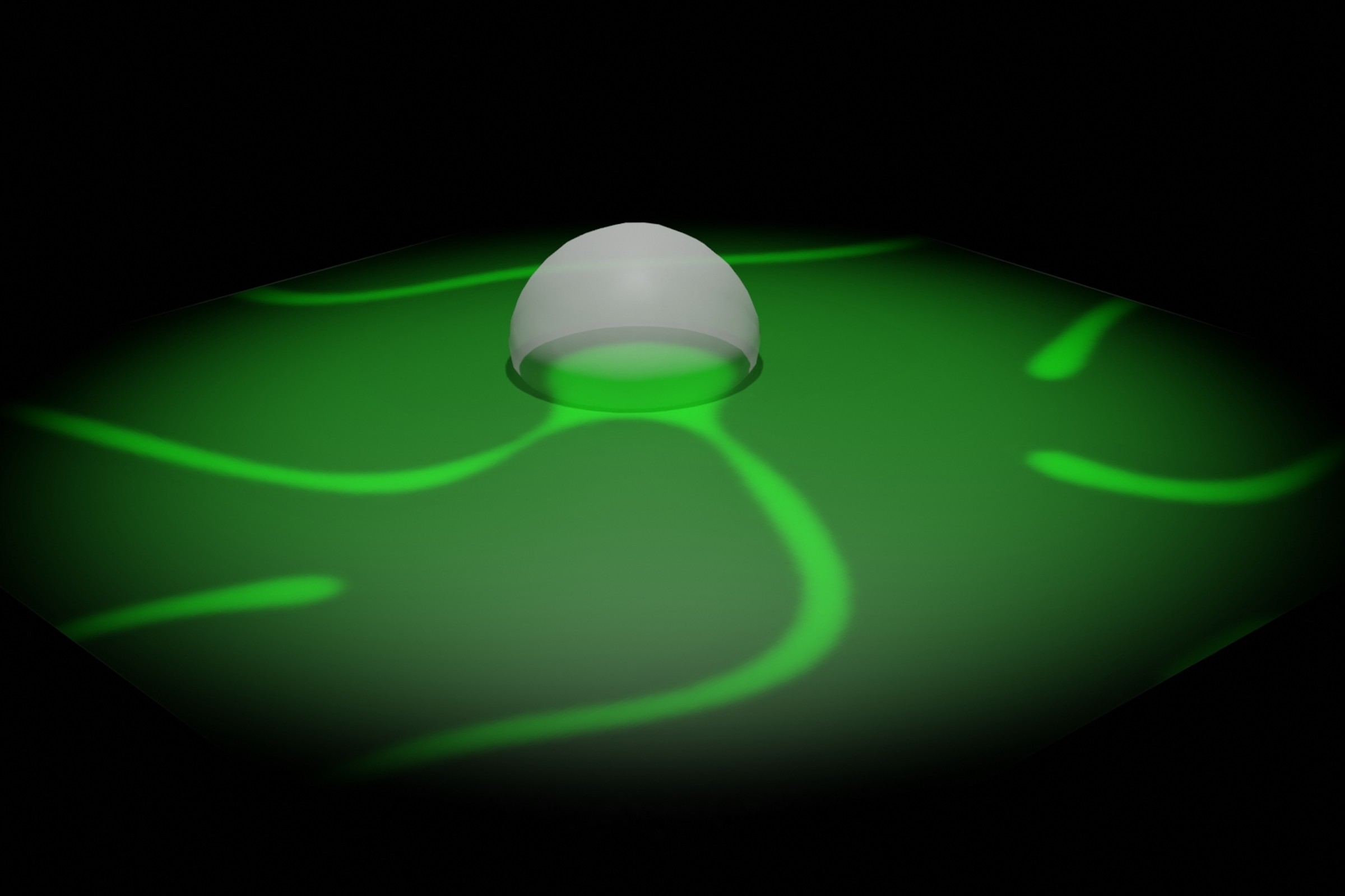
| Simulations showed that there are two possible mechanisms how the Min proteins interact with the liposomes. (Image: J. Willeke) | |
| Experiments in Schwille’s laboratory have shown that membrane-binding Min proteins in the artificial system arrange themselves asymmetrically around the vesicles and interact with them in such a way as to set them in motion. In the process, the proteins bind both to the supporting membrane and to the vesicles themselves. | |
| “The directed transport of large membrane vesicles is otherwise only found in higher cells, where complex motor proteins perform this task. To discover that small bacterial proteins are capable of something similar was a complete surprise,” observes Schwille. “It is currently unclear not only what exactly the protein molecules do at the membrane surface, but also for what purpose bacteria could need such a function.” | |
Two possible mechanisms |
|
| With the aid of theoretical analyses, Frey’s team identified two different mechanisms that could be behind the motion: “One possible mechanism is that the proteins on the supporting membrane interact with those on the vesicle surface somewhat like a zipper and form or dissolve molecular compounds in this way,” explains Frey. “If there are more proteins on one side than on the other, the zipper opens there, while it closes on the other side. The vesicle thus moves in the direction in which there are fewer proteins.” | |
| The second possible mechanism is that the membrane-bound proteins deform the vesicle membrane and alter its curvature. This change in shape then causes the forward motion. | |
| “Both mechanisms are possible in principle,” emphasizes Frey. “What we do know for certain, however, is that the protein patterns on the supporting membrane and on the vesicle cause the motion. This represents a big step forward on the road to artificial cells.” The authors are convinced that their system can serve as a modeling platform in the future for the development of artificial systems with life-like movements. |
| Source: Ludwig-Maximilians-Universitaet Muenchen (LMU) (Note: Content may be edited for style and length) |

