| Jan 03, 2024 | |
On-demand conformation of an artificial cytoskeleton |
|
| (Nanowerk News) Peptide nanotubes are tubular-shaped structures formed by the controlled stacking of cyclic peptide components. These hollow biomaterials show inner and outer faces, allowing the control over their properties. | |
| Led by Juan R. Granja, researchers from the Center for Research in Biological Chemistry and Molecular Materials (CiQUS) presented a novel kind of cyclic peptide that, when light-irradiated, induces the formation or desegregation of nanotubes on demand. At the appropriate wavelength, the peptide switches from a folded to a flat conformation. When the planar conformation is adopted, the peptide rings assemble to form tubular-shaped structures, whereas in the folded arrangement the peptides remain unassembled. | |
| The research has been published in Chem ("Photo-assembling cyclic peptides for dynamic light-driven peptide nanotubes"). | |
 |
|
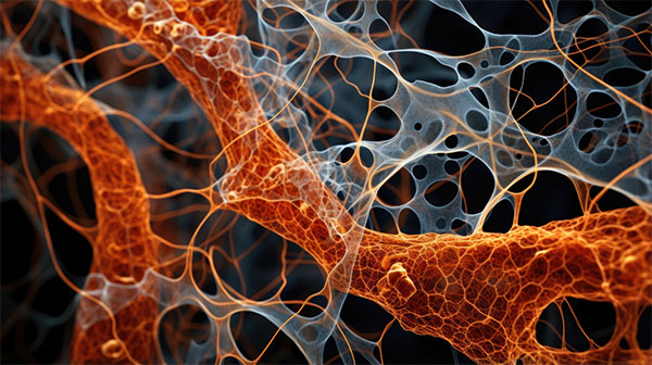
| Illustration of tubular-shaped peptide rings. (Image: CiQUS) | |
| A mesh of microtubules provide structure and shape to cells, and are key to perform their functions and divide. One of the main challenges towards cell biology is to emulate this fibers in order to construct an artificial cytoskeleton. | |
| To this end, Prof. Granja's group has been studying for years the properties of peptide nanotubes to create these synthetic meshes and thus control the molecular mechanisms underlying these biological processes. However, a simple model to simulate this vital component of cells requires the formation/disassembly process to occur with precise spatio-temporal control under physiological conditions, something that was not possible with peptide nanotubes at the time. | |
| Using light as an external stimulus, in this work CiQUS researchers synthesized the nanotubes inside water droplets under neutral conditions, thus simulating the physiological media present in the cell. Fibers formed rapidly on the contour of the water-in-oil droplets and induced their fusion. | |
| According to the authors, when the nanotubes are located at the interfase they provide the droplets with the ability to fuse with each other, a mechanism of great interest to simulate cell phagocytosis or to explore new systems for drug delivery. |
| Source: Center for Research in Biological Chemistry and Molecular Materials (Note: Content may be edited for style and length) |

