| Jul 03, 2012 |
First visualisation of the DNA double helix in water
|
|
(Nanowerk News) When Watson and Crick discovered the DNA double helix nearly sixty years ago, they based their structure on an averaged X-ray diffraction image of millions of DNA molecules. Though the double helix has become iconic for our molecular-scale understanding of life, thus far no-one has ever “seen” the double helix of an individual double-stranded DNA in its natural environment, i.e, salty water.
|
|
Dr Carl Leung and a team of international collaborators led by Dr Bart Hoogenboom at the London Centre for Nanotechnology (LCN) have now done just that (see paper in Nano Letters: "Atomic Force Microscopy with Nanoscale Cantilevers Resolves Different Structural Conformations of the DNA Double Helix").
|
 |
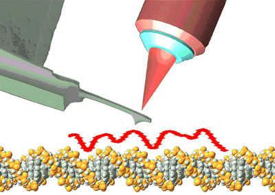
| A miniaturised cantilever tracing the contours of the DNA double helix, with its deflection detected by laser optics (not to scale).
|
|
To visualise DNA, they used a technique called atomic force microscopy (AFM) that detects the molecules by ‘feeling’ them, as a blind person would with a cane. Atomic force microscopy is known for its ability to achieve up to atomic resolution on flat surfaces, but often struggles to resolve more complex structures such as biological molecules. Imaging DNA, for instance, is analogous to using a cane to visualise a wriggling snake: not an easy task!
|
|
To resolve the double helix, the researchers miniaturised the cane (the “cantilever”) to approximately 10 microns length (an eighth of the width of a human hair) and nanometre-scale thickness. Next, they made it vibrate at sub-nanometre amplitudes and detected the proximity of the DNA via minute changes in the resonance frequency of the cantilever.
|
|
The resulting images show the two strands of the double helix twisting around the central axis of the molecule, in a clockwise, right-handed fashion, similar to a corkscrew entering a cork. The so-called major groove is clearly resolved, separating the turns of the double helix, as well as the minor groove that separates the two strands of the double helix. This level of detail sets these images apart from all other AFM measurements on DNA over the past two decades.
|
|
Interestingly, the reported method also enabled the researchers to identify and probe a surprising form of DNA that has a left-handed structure, as opposed to the canonical right-handed structure of Watson and Crick . In future this method could therefore be used to study structural deviations of the double helix, linked to genetic instability and hereditary disease.
|

