| Nov 23, 2012 |
Nanodiamonds as revealing feed for microorganisms |
| (Nanowerk News) Nanodiamond has emerged to be a new promising class of nanomaterial. It shows a stable Raman signal as well as a stable fluorescence without any photo-bleaching. In addition, its surface can be easily functionalized with various molecular and ionic groups followed by further conjugation with biomolecules of interest. Several studies have demonstrated that nanodiamond is biocompatible and that it is not cytotoxic for many cells. It can penetrate cells via endocytotic pathways, and accumulate inside. These properties make nanodiamond an interesting candidate for bioimaging and drug delivery purposes. |
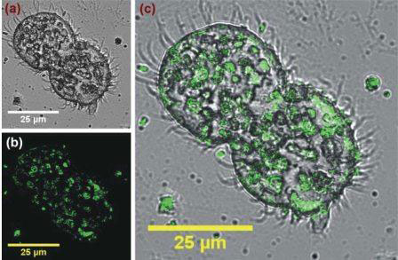 |
| Nanodiamond has great potential for biolabeling and drug delivery. Scientists tested the new material for intracellular in vivo imaging of protist microorganisms. |
| However, the knowledge on nanodiamond interactions with living organisms as well as with organs and tissues is still limited. Negative effects have been reported. To understand the nanodiamond interaction with living organism, the investigation of the effects on various protist microorganisms would be an important next step, but there have been very few studies so far. |
| A team led by Chia-Liang Cheng from National Dong Hwa University (Taiwan) now treated the protist microorganisms Paramecium caudatum and Tetrahymena thermophile as well as a murine macrophage cell line (RAW264.7) with nanodiamond particles in order to study the biocompatibility and the biolabeling properties of the nanomaterial ("Nanodiamond for intracellular imaging in the microorganisms in vivo"). |
| As fluorescence imaging of stained cells revealed, murine macrophages take up nanodiamond particles. Inside the cells, the tiny particles were found to be co-localized with lysosomes. The in vivo experiments with the two microorganism strains showed that their functioning was preserved during examination after treatment with nanodiamond particles. Yet, the growth rate was reduced. When comparing nanodiamond particles of 5 nm and 100 nm diameter, the smaller particles turned out to be more toxic, presumably due to their surface disordered carbons. In comparison to animal and human cell cultures, nanodiamond was found to be more toxic for microorganisms. During the experiments, it could be observed that the microorganisms “eat” the nanomaterial which is then transported into their food vacuoles. After a certain time, they excrete it. |
| In another experiment, the researchers labeled E. coli by adsorbing nanodiamond on the cell wall and fed the bacteria to T. thermophile. Via imaging of the nanodiamond particles, the position of E. coli could be detected inside the food vacuoles. |
| Source: By K. Maedefessel-Herrmann, Wiley |

