| Jan 07, 2013 |
Living cells behave as fluid-filled sponges |
| (Nanowerk News) Scientists from the London Centre for Nanotechnology at UCL have shown that animal cells behave according to the theory of ‘poroelasticity’ when mechanically stimulated in a way similar to that experienced in organs within the body. The results indicate that the rate of cell deformation in response to mechanical stress is limited by how quickly water can redistribute within the cell interior. |
| Poroelasticity was originally formulated to describe the behaviour of water-saturated soils and has important applications in the fields of rock engineering and petro-physics. It is commonly used in the petroleum industry. Poroelastic models describe cells as being analogous to fluid-filled sponges. Indeed, cells are constituted of a sponge-like porous elastic matrix (comprising the cytoskeleton, organelles, and macromolecules) bathed in an interstitial fluid (the cytosol). In this analogy, the rate at which the fluid-filled sponge can be deformed is limited by how fast internal water can redistribute within the sponge in response to deformation. This rate is dictated by three parameters: the stiffness of the sponge matrix, the size of the pores within the sponge matrix, and the viscosity of the interstitial fluid. |
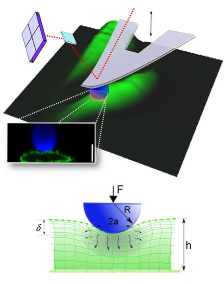 |
| Atomic Force Microscopy (AFM) microindentation enables investigation of the dynamic mechanical properties of living cells. Schematic of an AFM cantilever with a spherical tip indenting a cell. The image of the cells (green) is a 3D reconstruction from AFM topographical data. A confocal microscopy image shows the cell profile as the cell is deformed by a spherical bead (blue). The cell membrane is revealed using a fluorescent protein (shown in green) and a fluorescent bead is attached to the cantilever (in blue). Scale bar =10 µm. Below the profile image, a schematic diagram illustrates the indentation of a fluid-filled sponge (poroelastic material). Rapid indentation of the cellular material induces pressurisation of the interstitial fluid leading to its movement out of the compressed area. As the fluid redistributes, the pore pressure diminishes and the reaction force decreases. |
| To study cellular responses, LCN scientists used cell-sized levers to apply rapid well-controlled deformations on the cell surface and monitored the temporal response of cells to these deformations. Close examination of the experimental results revealed that the rate of cellular deformation was limited by how rapidly water could redistribute within the cell interior. Experimental measurements indicated that this sponge-like behaviour of cells likely occurs during normal function of organs such as the lungs and the cardiovascular system. |
| Emad Moeendarbary, lead author of the paper ("The cytoplasm of living cells behaves as a poroelastic material") from the LCN said: "In the cardiovascular system, some tissues encounter extreme mechanical conditions. Heart valves can typically withstand seven-fold increases in their length in less than one second. The poroelastic nature of cells may allow them to behave similarly to shock absorbers when exposed to these extreme mechanical conditions." |
| To experimentally verify the fluid-sponge model, researchers manipulated the size of the cellular pores using chemical and genetic tools and showed that the rate of cellular deformation was affected by the pore size, as suggested by the theory of poroelasticity. |
| Guillaume Charras, senior co-author of the paper from the LCN said: “Cells can detect the mechanical forces they are subjected to and modify their behaviour accordingly. How changes in the mechanical environment are converted into biochemical information that the cell can interpret remains unknown. A better understanding of the physics of the cellular material is a first step towards formulating possible mechanisms through which this could occur.” |
| Source: London Centre for Nanotechnology |

