| Feb 04, 2013 |
Ultrafast chemical imaging lights the way to monolayer and nanometer spatial resolution |
| (Nanowerk News) Since the 19th century, microscopy and spectroscopy methods have illuminated many aspects of chemistry and physics, from defining atomic spectra to bringing clarity to Einstein’s photoelectric effect. |
| Now, in the early 21st century, chemical images generated via high-resolution spatiotemporal measurements combined with spectroscopy are edging us closer to a scientific dream: to visualize single-molecule or atomic scales in situ and in real time. Molecular research in organic photovoltaics, polymers, macro/supra-molecular self-assembly, biomembranes, proteins, and generally matter organized to form nanoscale molecular structures all could benefit from this burgeoning ultrafast, femtosecond nano-imaging capability. |
| For Markus Raschke, a professor at the University of Colorado at Boulder and current EMSL Wiley Research Fellow, the evolution toward achieving this scientific innovation is driven by his long-term interest in ultrahigh spatial resolution optical imaging and spectroscopy. This interest initially brought him to EMSL as a user and has since led to a nearly four-year collaboration that has EMSL standing at the precipice of delivering imaging capabilities with near single-molecule sensitivity. |
| The ‘Tip’ping Point |
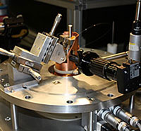 The s-SNOM, which became a long-term project that initially stemmed from a Scientific Partner Proposal, is housed among EMSL’s multifaceted Microscopy Capability instrument suite. Raschke and his colleagues initially used EMSL’s microscopy capabilities to demonstrate plasmonic nanofocusing using an optical antenna concept. The method employed a conical gold tip and short-pulse excitation to facilitate background-free near-field imaging via scattering-type scanning near-field optical microscopy, or s-SNOM. The combination also affords nanofocusing of femtosecond pulses and optical control on the nanoscale. It opened the door to nanoscale ultrafast spectroscopy that could depict matter amid its real time and length scales simultaneously, as well as to control a single quantum excitation with that unique light source “at the tip of a needle,” according to Raschke. “We wanted to design a nanoscale light source,” Raschke explained. “We sought different avenues to get to this goal and achieve this for different wavelengths and timescales. Fabricating these tips, which act as special conical waveguides, provide for a highly confined light source, where the optical field energy is compressed into a very, very small volume at its apex.” While the ultrafast spectroscopy aspect was not Raschke’s initial goal, the success achieved in that effort ("Light on the Tip of a Needle: Plasmonic Nanofocusing for Spectroscopy on the Nanoscale") offered EMSL an incredible opportunity to enhance the understanding of chemistry on surfaces and interfaces – where environmental, catalytic, and biological interactions occur and chemistry happens – by way of its Scientific Partner Proposal process. |
| Building a Partnership |
| Interested partners, like Raschke and his colleagues, submit proposals via the EMSL User Portal to team with EMSL staff and enhance existing capabilities or develop new ones. In this case, EMSL’s American Recovery and Reinvestment Act funding facilitated the development of the infrared, or IR, scattering type scanning near-field microscope, which was initially housed at Raschke’s laboratory as he and his team built, tested, and optimized the new capability. Earlier this year, the customized IR s-SNOM microscope was moved into its home at EMSL, where Raschke, along with EMSL scientist Ian Craig, still are at work honing its development and applications. |
| “At EMSL, we’ve long been focusing on technology dealing with improved spatiotemporal resolution that lets us look at chemistry in real-world conditions,” said David Koppenaal, EMSL’s chief technology officer. “This is a unique capability that will provide high-resolution molecular information at the nanoscale. And, it complements several microscopy capabilities we have here already.” |
| According to Raschke, EMSL’s scientific partnering mechanism also is a prime example of interdisciplinary and collaborative science, the kind of investment that motivates scientists and promotes new scientific frontiers. Coming from the academic side, he knows how valuable this interaction can be toward achieving tangible innovation. |
| “We did not have the resources or infrastructure to create an instrument with these wonderful capabilities at the academic level,” Raschke noted. “Partnering with EMSL put the best of both worlds together: the dynamics and enthusiasm at a university and the resources and capability at EMSL. We all want the best science.” |
| The Innovators |
| After demonstrating s-SNOM’s potential to extend IR spectroscopy into the nanometer scale based on their optical antenna concept, Raschke and his colleagues partnered with EMSL to take on the challenge of improving its spectroscopic sensitivity. |
| “It is well known that you can see a single molecule using an atomic force or scanning tunneling microscope, but you do not get spectroscopic detail – and those techniques, albeit exquisitely sensitive, are too slow to get the internal dynamics,” Raschke said. |
| “Lasers give you high spectral resolution, and pulsed lasers tell you about dynamics in matter,” he continued. “But, the spatial resolution is limited for looking at the finer details of the molecular composition. What we did was to really combine the sensitivity and spatial resolution of scanning probe microscopy with ultrafast laser spectroscopy to get the best of both worlds.” |
| By combining both tip and substrate enhancement gleaned from their initial work with optical antennas and molecular Raman spectroscopy and improved signal-to-noise ratio from high-spectral irradiance IR pump excitation, Raschke and his colleagues imaged a self-assembled monolayer, or SAM, made from 16-mercaptohexadecanoic acid, a compound used in self-assembly to produce hydrophilic SAMs, on a gold surface. They were able to obtain 25-nm spatial resolution using their IR s-SNOM technique and could spectroscopically determine the chemical identity of the surface molecules. Most significantly, they broke a record in spectral sensitivity and contrast, getting signal from only ~100 molecular vibrations – almost nine orders of magnitude more sensitive than conventional IR spectroscopy. |
| “This paves the way toward single-molecule IR spectroscopy,” Raschke said ("Pushing the Sample-Size Limit of Infrared Vibrational Nanospectroscopy: From Monolayer toward Single Molecule Sensitivity"). “We’ve shown you can get a signal. We’re looking at 100 molecules when before I had colleagues who didn’t think one could even get a signal from 1 million molecules.” |
| As the senior scientist and collaborator throughout this effort, Raschke continues to publish papers as he refines the IR s-SNOM, seeking means to improve its capability. He also welcomes its evolution as another unique instrument that EMSL offers for broad use to the scientific community. His leadership role is one he fully expects, and is excited, to continue for years to come. |
| “If you can see it on a single molecular monolayer, you can see it on anything, really,” Raschke said. “We are seeing how light interacts with matter on the clock of nature. We look at the motion of electrons and atoms in real time. We have seen collections of atoms do this. But, you need many to get a signal. Now, we are getting to where we can see the atomic motion of individuals. |
| “One hundred molecules is an important number. This is where atoms become a family. We are getting down to the homogenous ensemble, seeing the heartbeat of matter,” he added. |
| Source: Environmental Molecular Sciences Laboratory |

