| Mar 08, 2013 |
Rapid cancer detection on a chip |
| (Nanowerk News) Early detection is vital for the effective treatment of cancer. In many cases, tell-tale biomarkers are present in the bloodstream long before outward symptoms become apparent. Early-stage cancers, for example, release tiny quantities of biomolecules called microRNAs into the blood. The development of an inexpensive and rapid point-of-care diagnostic test capable of spotting such early biomarkers of disease could therefore save many lives. A research team in Japan working on developing such a test has now produced their most sensitive microRNA detector yet ("Rapid and Sensitive MicroRNA Detection with Laminar Flow-Assisted Dendritic Amplification on Power-Free Microfluidic Chip"). |
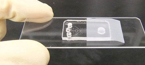 |
| A self-powered microfluidic chip for cancer biomarker detection. The sample and two fluorescence amplification reagents are added to the three inlet ports. The presence of cancer biomarkers can be detected by fluorescence in the main microfluidic channel. |
| The test developed by Kazuo Hosokawa and colleagues at the RIKEN Advanced Science Institute is a self-powered microfluidic chip (Fig. 1) that can perform an analysis for cancer-specific microRNAs in a drop of patient blood in as little as 20 minutes. If enough of the target microRNA is present, the chip produces a fluorescence signal that can be detected using a fluorescence microscope ("Rapid microRNA detection using power-free microfluidic chip: coaxial stacking effect enhances the sandwich hybridization"). |
| The team’s microfluidic chip is inexpensive to make and relies on an internal pressure gradient to pump the sample through the microchannels, thus eliminating the need for an external power supply—features that make the system highly suitable for practical point-of-care disease diagnosis. Previous versions of the chip, however, could only detect microRNA at concentrations far above those required for early cancer detection. |
| In their latest work, Hosokawa and co-workers increased the chip’s sensitivity by boosting the intensity of fluorescence generated by a positive test. The original chip worked by immobilizing target microRNA on probe DNA in the main microchannel, where each bound site produced a fluorescent signal. In the new chips, the researchers added a fluorescence amplification process that involves passing two amplification reagents over the immobilized microRNA. The reagents—a fluorescent tag and a branched linker—bind to immobilized microRNA to form tree-like dendritic structures that amplify the fluorescence signal by up to 1,000 times. Using this amplification process, the researchers were able to improve the sensitivity of the device to a level approaching that required for early cancer detection. |
| The next step for the team will be to further simplify the device by eliminating the need for a fluorescence microscope, which will involve replacing the fluorescent tags with some other form of marker. “That is a very important direction for future development,” says Hosokawa. “We are planning the use of different labeling materials instead of the fluorescent dye, such as gold particles, which would enable naked-eye detection,” he says. The team is also working to improve the sensitivity of the technique. |
| Source: RIKEN |

