| Mar 25, 2013 |
Researchers use 'Spinostics' to detect cancer biomarkers (w/video)
|
|
(Nanowerk News) A new biosensing assay which can specifically and rapidly detect colorectal cancer biomarkers in solution has been developed by researchers at the London Centre for Nanotechnology and other UCL departments, introducing the concept of “spinostics”.
|
|
Reported in Nature Scientific Reports ("Homogeneous antibody fragment conjugation by disulfide bridging introduces ‘spinostics’") this week, the study describes the addition of a “spin label” to an antibody fragment, which can then be detected using electron spin resonance (ESR) and subsequently used to determine the presence of certain cancer biomarkers.
|
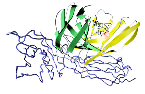 |
| The complex formed between the antibody fragment with the label (green and yellow) and the antigen (blue wire frame). The cartoon is a crystallographic representation – only a schematic.
|
|
This research has potential to provide a wide range of off-the-shelf biochemical tests, which use antibodies to detect the presence of biomolecules, revolutionising the in vitro diagnostics field.
|
|
The study describes the modification of the antibody fragment (anti-CEA sscFv) through its disulfide bond by adding a spin label, a nitroxide molecule that has an unpaired electron, which can attach itself to a biomolecule.
|
|
When this modified antibody fragment binds to the colorectal cancer biomarker, carcinoembryonic antigen (CEA), the tumbling motion of the fragment is slowed. These changes are reported by the attached spin label and can be detected using ESR.
|
|
Biomarkers are biological molecules found in blood or other body tissues that are a sign or signature of a disease or condition. The carcinoembryonic antigen (CEA), a protein involved in cell adhesion, is a biomarker of colorectal cancer and, if present in the blood, is an indication that the disease is present.
|
|
One of the advantages of this technique was that the researchers were able to measure CEA concentrations in complex media, such as whole human blood without the need to pre-treat the blood samples e.g. remove the red blood cells.
|
|
|
|
Another major advantage is the specificity and rapidity of binding, complete within 15 minutes.
|
|
This study shows that there is broad potential for antibody-based in-solution diagnostic methods using ESR or ‘spinostics’ and that this technology offers a platform for the development of new tools to study biological systems and for application in diagnostics and biotechnology.
|
|
Vishal Sanchania, one of the leading authors from the London Centre for Nanotechnology, commented “With the advent of ‘spinostics’ we envisage the generation of a vast library of ESR in vitro diagnostics (IVD) assays, with great future promise for immunoassays.”
|
|
He added “Currently, IVD are one of the only ways to detect and quantify diseases. Hence, there is an ever increasing need for new IVD technologies to provide more efficient and rapid detection methods that require little time from the diagnostician. ‘Spinostics’ offers some great advantages, including simplicity and readily interpretable quantitative outputs, over currently leading immunoassay technologies. “
|

