Optical tweezers explained
Discover the evolution of optical tweezers, a powerful tool used to manipulate particles at the microscopic scale. Explore their history, advancements, and wide-ranging applications in nanotechnology, biological research, and materials science, including the incorporation of graphene and holographic optical tweezers.
Optical tweezers have come a long way since their inception, with numerous milestones and key developments marking their journey. Understanding the history of optical tweezers offers valuable insight into their evolution and their impact on various scientific fields.
The foundations of optical tweezers, also known as optical traps, can be traced back to the early 20th century when the idea of radiation pressure and the momentum carried by light was first explored. In 1901, the renowned physicist Max Planck introduced the concept of quantized energy levels in light, which ultimately led to the realization that light could exert pressure on particles. However, it took several decades before these principles were applied to manipulate microscopic particles.
The history of optical tweezers
The concept of optical trapping was first proposed in the 1960s and 1970s by Arthur Ashkin, a scientist at Bell Laboratories. In 1970, Ashkin demonstrated that a focused laser beam could trap and manipulate dielectric particles, laying the groundwork for future optical tweezers (see the original 1970 paper: "Acceleration and Trapping of Particles by Radiation Pressure"). However, it wasn't until 1986 when Ashkin, along with his colleagues Steven Chu and John Bjorkholm, developed the first optical tweezer system capable of trapping and manipulating individual particles. Their design used a high numerical aperture objective lens to focus the laser beam, creating the intense light spot necessary for optical trapping.
In 2018, Arthur Ashkin was awarded half of the Nobel Prize in Physics 'for groundbreaking inventions in the field of laser physics'.
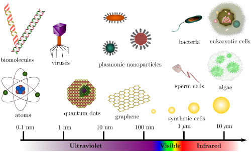
Optical tweezers have become a powerful tool in nanotechnology to use light to manipulate microscopic objects as small as a single atom. They have been utilized for capturing a range of objects, including dielectric spheres, viruses, bacteria, living cells, organelles, small metal particles, and even DNA strands. These tweezers have numerous applications, such as confining and arranging objects for cell sorting, monitoring movement of bacteria, measuring small forces, and manipulating larger structures like cell membranes.
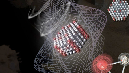
More recently, researchers even have developed a new way to study the intricate dynamics within living cells by using optically trapped nanodiamond particles inside leukemia cells as intracellular sensors and then measured magnetic noise within the cell. This new technique allows for highly precise measurements of temperature and magnetic fields inside living cells, which can help researchers study cellular properties and detect harmful molecules called free radicals. This breakthrough may eventually lead to better diagnosis and treatment of diseases like cancer and neurodegenerative disorders.
Working principle of optical tweezers
Optical traps use one or more focused laser beams to hold and manipulate tiny particles. Imagine a magnifying glass concentrating sunlight to a single point; that's similar to how the laser beam works in optical tweezers. The particles being manipulated have to let light pass through them differently than the surrounding material, like a glass bead in water.
Two types of forces work together to trap the particle near the laser's focal point. First, there's the scattering force, which comes from the pressure of the light beam and pushes the particle in the same direction as the beam. Second, there's the gradient force, which attracts the particle towards the area with the highest light intensity (the focal point). These forces combine to hold the particle in place, allowing scientists to study and manipulate it.
These are sophisticated and costly instruments, typically custom-built by adapting commercial optical microscopes with extensive modifications. Integrating multiple lasers into the microscope presents a significant challenge. To minimize photo damage to biological samples while maintaining high trapping stiffness, high-power infrared laser beams are often employed. The optical trap's precise maneuvering is achieved using lenses, mirrors, and acousto/electro-optical devices that can be computer-controlled. The diagram below illustrates the complexity of such systems, which necessitate expertise in microscopy, optics, and laser techniques.

Development of holographic optical tweezers
In the early 2000s, researchers developed holographic optical tweezers, a groundbreaking technology that uses computer-generated holograms to simultaneously create multiple traps for manipulating hundreds of particles at a time. This innovation greatly expanded the capabilities of traditional optical tweezers, enabling scientists to study more complex systems by handling multiple particles at once.
Holographic optical tweezers create versatile 3D configurations of single-beam optical traps, which are used to manipulate mesoscopic objects. They achieve this precision by employing beam-splitting, mode-forming, and adaptive wavefront correction techniques. These advanced tweezers can apply highly controlled forces and torques on objects ranging in size from nanometers to hundreds of micrometers.
With their nanometer-scale spatial resolution and real-time reconfigurability, holographic optical traps provide unparalleled access to the microscopic world. This technology has found a wide range of applications in fundamental research, manufacturing, and materials processing, further demonstrating its impact and potential.
Invention of opto-thermoelectric tweezers (OTET) and the incorporation of graphene
Thermal effects caused by laser beams in the focus area pose challenges like potential damage to biological samples and reduced particle-trapping accuracy due to stronger Brownian motion. To address these issues, various optical tweezer techniques have been developed, but they often don't fully satisfy the critical demands.
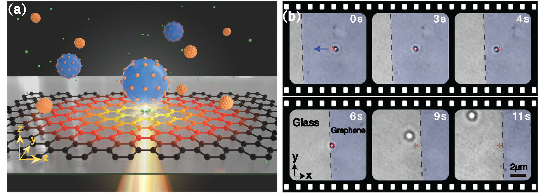
Opto-thermoelectric tweezers (OTET), developed in 2018, are promising as they require significantly lower laser powers compared to traditional optical tweezers. Researchers at Shenzhen University have further enhanced the performance of OTET by replacing the gold film with graphene, resulting in a broader working wavelength range and a larger trapping area compared to gold-film-based OTET. The graphene-based OTET decreased the damage caused to biological samples and extended the working wavelength range, making it suitable for a wide variety of research applications.
Advance in biomedical research
Researchers have also discovered a new technique that overcomes a fundamental technical challenge in optical tweezers, allowing them to manipulate particles with the same refractive properties as their background environment. This breakthrough has significant potential in medicine, as it enables the manipulation and measurement of microscopic objects inside cells, such as strands of DNA or intracellular enzymes, with greater precision and less invasiveness.
The researchers developed a unique method to control the refractive properties and luminescence of nanoparticles by doping nanocrystals with rare-earth metal ions, leading to much lower energy levels and 30 times increased efficiency compared to traditional methods. The heat generated by this new method was also negligible compared to older methods, offering a number of advantages.
Capturing nanoplastics in tap water with light
A practical application of optical traps is a real-time detection system for nanoplastics using electro-photonic tweezers and metal nanoparticles. Researchers combined electrical nanoparticle capture with real-time Raman spectroscopy, which analyzes energy differences between light frequencies, to detect nanoplastics as small as 30 nm. This method also reduced the time required for the entire process, including collection, separation, and analysis, from one day to just a few seconds. The researchers believe this new technique can be used to measure microplastic concentrations in various water resources, and it may contribute to securing clean water supplies.
Fluorescence color control
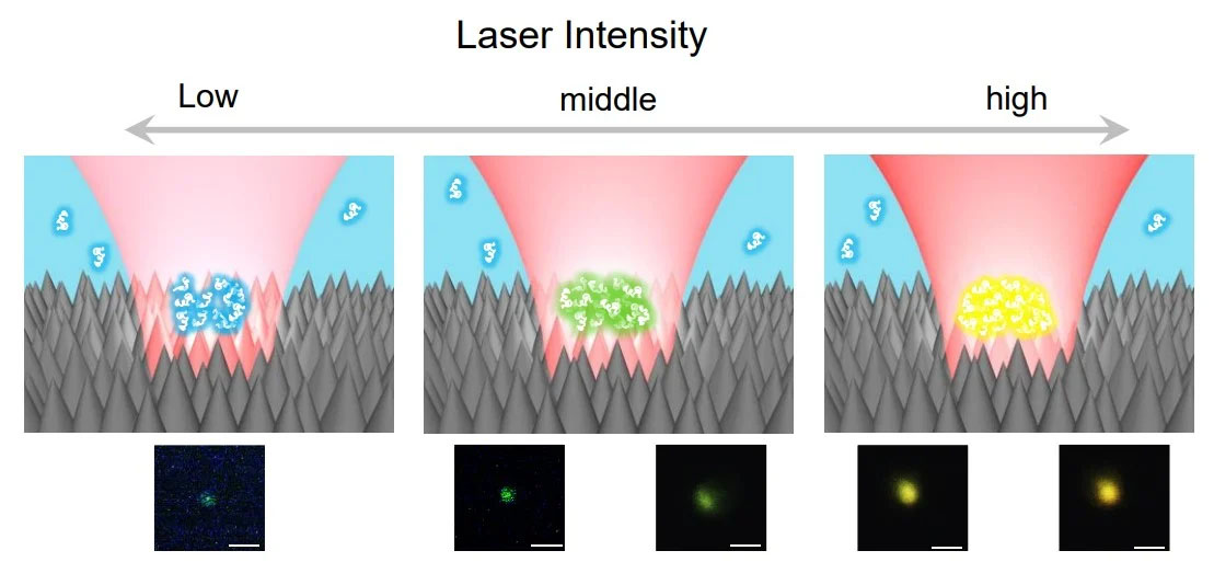
Scientists have developed an optical trapping technique using nanotextured black silicon that can trap and manipulate polymer chains. They used optical tweezers to remotely control the fluorescence color emitted by adjusting the laser intensity. This technology can create a range of colors from blue to orange, covering most of the RGB spectrum.
One big stumbling block in the field of photonics is that of color control. Previously, controlling color required altering the chemical structure or concentration of the emitter, which needed direct contact and limited its application. Optical tweezers enable remote and quick color control, suitable for microscopic spaces and closed systems.
From a low intensity blue to high intensity orange, this reversible and fully remote technology can almost reach the entire RGB spectrum.
This technology is still in the early stages, but it has potential applications in ultraviolet and near-infrared regions, as well as in micromachine components and intracellular bioimaging.
Conclusion
The history of optical tweezers is marked by numerous milestones and key developments that have shaped the technology into the powerful tool it is today. From their early beginnings to their current applications in nanotechnology, biological research, and materials science, optical tweezers continue to revolutionize the way we study and manipulate particles at the microscopic scale. The incorporation of graphene into opto-thermoelectric tweezers has shown the potential for further advancements in the field, such as combining the system with microfluidic technology for on-chip applications like cell sorting and investigating cell-to-cell interactions. Additionally, the exploration of other two-dimensional materials and artificial metamaterials may lead to even more enhancements and expanded functions of optical tweezers.
As optical tweezers continue to evolve and integrate with other cutting-edge technologies, they are expected to play an increasingly significant role in numerous scientific disciplines. Researchers are continually pushing the boundaries of what is possible with optical tweezers, enabling new discoveries and breakthroughs in areas ranging from medicine and biology to materials science and nanotechnology. The future of optical tweezers is undoubtedly bright, and as the technology continues to advance, its impact on science and technology is only expected to grow.
