| Posted: Dec 16, 2009 | |
Atomic Force Microscopy and combined optical techniques |
|
| (Nanowerk Spotlight – Application Note) This Application Note briefly describes the basics of both optical and atomic force microscopy, followed by a discussion of some of the technical challenges of integrating these two distinct imaging modalities. In certain cases, the benefits and disadvantages of different approaches to design and integration are discussed. Lastly, a few examples of successful application of these combined imaging modalities are presented. | |
| Introduction | |
| Since its invention in 1986 by Binnig, Quate, and Gerber ("Atomic Force Microscope"), the atomic force microscope (AFM) has become an indispensable tool for investigators in the physical, materials, and biological sciences. The AFM quickly gained acceptance in these fields due to its ability to capture topographical maps of surfaces in either air or liquid with sub-angstrom (in Z) and nanometer (in XY) resolution. Further, the ability of the AFM to measure a variety of forces with pico-Newton precision quickly led to measurements of single-molecule and intra-/intermolecular forces. Force measurements were also extended to the measurement of the elasticity of samples such as living cells and polymers that are typically too soft to measure precisely with traditional instrumented nanoindentation techniques. Integral to the success of AFM-based techniques is the relatively simple and label-free preparation of most samples when compared to other ultra-microscopical techniques. | |
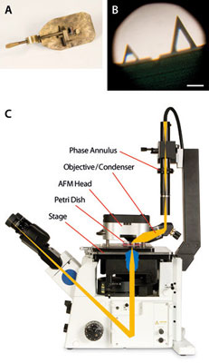 |
Figure 1: Early optical microscopes (A) were fairly simple devices utilizing ball lenses and samples mounted with beeswax. Magnifications upwards of ∼250X were possible; enough to easily resolve modern-day AFM cantilevers (B, scale bar = 100µm). Inverted optical microscopes can be coupled to scanning probe microscopes without restricting optical resolution (C) and providing top-down optical access (schematic in C, shown: MFP-3D-BIO™ AFM from Asylum Research integrated with inverted optical microscope). |
| Today, extensive imaging modalities have been implemented on the AFM under the umbrella of scanning probe microscopy (SPM). In addition to topographical imaging, SPM has been used to measure magnetic fields, friction gradients, potentials, capacitance, current flow, piezo response, and temperature (to name a few) across a diverse array of samples. Wider commercial availability of user-friendly instrumentation has put the AFM into the hands of more researchers, not only pushing the boundaries of its application in particular fields, but also bringing together scientists at the interfaces between disciplines. | |
| An exciting and promising area of growth for AFM has been in its combination with optical microscopy. Although new optical techniques developed in the past few years have begun to push traditional limits, the lateral and axial resolution of optical microscopes are typically limited by the optical elements in the microscope, as well as the diffraction limit of light. However, its ability to image through the entire depth of certain samples with chemical specificity using a plethora of label-conjugated markers allows researchers to identify specific structures or molecules within a dynamic event. Coupled with AFM’s ability to measure high-resolution topographical images, forces, and/or elasticity on a sample, a more complete understanding of structure-function relationships can be elucidated with a combined AFM/optical system. While the two imaging modalities have been used in combinational studies for over a decade, significant challenges of direct correlation of the two data sets have existed primarily due to the scaling differences between the two data sets. Recent developments in software now allow for user-friendly and intuitive routines for direct overlay and comparison between the two data sets. Further, various optical techniques are now being used to modify or stimulate samples of interest in concert with AFM measurements, and vice-versa. Indeed, AFM researchers find themselves in a diverse, multi-interfacial area of microscopy, made even more powerful by combining AFM with optical microscopy. | |
| Microscopy Basics | |
| While the first commercial AFMs were produced in the late 1980s, the origins of optical microscopy are much less clear, but are thought to lay with simple magnifying glasses in the mid-9th century with further developments in the 16th century. However, it wasn’t until the 17th century that history records scientific observations made with simple and compound microscopes, most notably in the field of biology by Hooke and van Leeuwenhoek (Figure 1A,B). Despite this long history, the most exciting time in optical microscopy has arguably been the past 100 years or so, as diffraction-limited optics, chemically-specific stains, and fluorescent markers and indicators have become widely available. In most modern applications, optical microscopy resolution is on the order of 200-300nm in X and Y, and 500nm in Z. | |
| The AFM (Figure 1C) uses a microfabricated cantilever made of silicon or silicon nitride with a sharp tip that physically touches the surface of interest. The cantilever raster-scans the sample while its deflection or oscillation amplitude is measured. | |
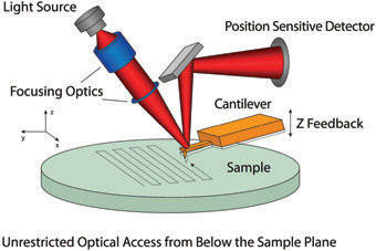 |
Figure 2: Schematic depicting the optical lever detection system used in AFMs. Light is reflected off of the back of a cantilever and read on a detector as the cantilever/probe is raster scanned to measure the topography of the sample. Most of the AFM instrumentation can be located above the sample, allowing for unrestricted optical access from below. |
| These measurements are performed with an optical tracking system that uses a segmented photodetector to track the reflection of a laser or superluminescent diode (SLD) off the back of the cantilever (Figure 2). Detected changes in cantilever deflection or oscillation are corrected to a setpoint value by actuating the cantilever in Z via a feedback-controlled piezo. These correction voltages sent to the Z piezo are recorded and correlated to a voltage-distance calibration factor in order to determine the height at a given XY coordinate. Because piezos suffer from nonlinearities due to hysteresis, creep, drift, and aging effects, most modern AFMs incorporate sensors that can linearize and correctly measure actual piezo actuation in XYZ. While a variety of sensors are available, the highest performance typically comes from linear variable differential transformers (LVDTs) because of their high linearity and low noise, which result in accurate tip and sample positioning to 0.06-0.6 nanometers. Additionally, the tip and the sample can be mounted on flexure stages that further linearize actuation. | |
| One of the great benefits of AFM is its ability to measure at multiple spatial scales. AFM resolution in XY is limited by the size of the tip, and is typically on the order of a few nanometers, while the upper measurement limit is on the order of 100 microns. Resolution in Z, however, is limited by electronic and thermal noise and is on the order of an Angström, with an upper measurement range that can be several tens of microns. In addition to measuring the physical topography of samples, the AFM cantilever can be used to measure forces such as adhesion, deformation, and sample elasticity by measuring the deflection of the cantilever versus tip-sample separation and applying simple spring mechanical models. With this approach, forces in the picoNewton range can be readily discriminated. | |
   |
|
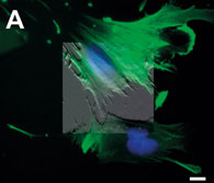
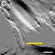
| Figure 3: AFM data can be overlaid and blended (A) to desired intensity (50% transparency shown) onto multiple color fluorescence data in order to correlate structures of interest such as the fluorescently-stained nucleus (blue). Detail (B) of the scanned region shows that fluorescently-labeled actin filaments (green) are not detectable at the border between two different cells with the optical microscope. Conversely, the AFM (C) reveals the presence of small cellular processes (arrow) connecting the two cells. Scale bars 10µm. | |
| A combined AFM/optical microscope is an excellent instrument for characterizing various samples. Optical microscopy’s chemical specificity and ability to image live processes within the depth of a sample is well complemented by the higher resolution capability of the AFM. For example, a popular technique for identifying internal components in cells utilizes multiple fluorescent markers that bind specifically to molecules of interest (Figure 3A). Overlaying the AFM data directly onto the optical data can allow for correlation, while the higher resolution of the AFM can resolve structures that are not composed of the target molecules for fluorescence, or structures that are too small or weakly labeled (Figure 3B,C). Integrating these two technologies is challenging, and different design criteria must be met to ensure success. | |
| Technical Design: Challenges and Solutions | |
| Various technical challenges exist when integrating optical and atomic force microscopes. Optically interfaced AFMs require a robust, custom-made stage to both support the instrument and minimize mechanical noise (Figure 1C). Typically, these stages must also be designed so as not to limit the movement or selection of optical microscope objectives, as well as to curtail the risk of mechanical interference between the stage and the optical microscope. Further, the design of the AFM components and stage must accommodate the various piezos, flexures, and sensors discussed above while minimizing the mechanical loop between the tip and the sample, as the susceptibility of the AFM to noise and thermal drift increases with the size of this loop. | |
| Because the AFM uses a laser or SLD to track the position of the cantilever, there must be several considerations in the design of the optical lever in order to minimize interference and crosstalk to the desired optical data. Some of the earliest commercial instruments utilized a red laser diode for optical lever tracking. These diodes were problematic for AFM in general, as they would readily couple optical interference fringes into AFM images and force curves taken on reflective samples. Additionally, the visible red diodes would be seen in images taken on the optical microscope, concomitantly preventing the detection of fluorophores that excite or emit within the wavelength range of the diode. Interference fringes coupled into AFM data were greatly attenuated by the introduction of low-coherence SLDs that emit infrared (IR) wavelengths. Though SLDs greatly reduce AFM data noise and reduce the amount of light emitted at visible wavelengths, the nature of SLDs does result in a faint emission in visible wavelengths that can interfere with highly sensitive optical measurements. Addition of a narrowband filter at the SLD source eliminates this interference, though many commercial AFMs omit such a filter due to the cost or the difficulty of incorporation into the optical lever design. And although many scientific-grade CCD cameras that are used to record images with the optical microscope are sensitive to wavelengths in the IR, addition of a well-designed filter under the microscope objective can block these signals from saturating the camera electronics. | |
| A further design difficulty presents itself with the incorporation of top-down optical access into AFM designs. While several AFM designs do not limit optical access below the sample (resulting in the ability to place the AFM on inverted optical microscopes), certain samples may be opaque or may require mounting on opaque sample holders that prevent the use of the inverted microscope optics. These issues are becoming more prevalent since opaque samples such as polymers, ceramics, and silicon-fabricated devices are garnering more interest across the materials, engineering, and biological sciences. In order to achieve quantitative data collection, high linearity, and low noise performance, the various components of the AFM must be mounted directly above the optical lever, which limits top-down optical access. With this in mind, it would seem that AFM designs would be forced to trade off data quality with optical access. | |
| One common solution to this challenge is to incorporate a large access hole in the AFM head, directly above the optical lever. While this design does allow for optical access directly through the top of the AFM head and the utilization of some of the transmitted light condensers from the optical microscope manufacturers, it also compromises the mechanical design of the AFM head and the accuracy of the optical lever, preventing the collection of truly quantitative force data. | |
| In some designs, either ancillary cameras and optics must be added next to the instrument or an entirely different base must be utilized for visualization and documentation of the top-down view for opaque samples. These measures complicate experimental design due to space restrictions in the necessary acoustic isolation equipment or in the difficulty in mechanical reproducibility when moving the AFM to different equipment. Other designs (Figure 1C) maintain the various components of the AFM head directly above the tip in order to preserve high-quality quantitative data collection, and incorporate a series of mirrors and high-quality objective lenses to provide top-down optical access through a customized optical microscope condenser (Figure 1C). While more costly, the added advantage of this design is that the objective lens can be used as both a condenser for transmitted light techniques, such as brightfield and optical phase contrast, as well as a viewing element for optical lever alignment and region-of-interest identification on opaque samples without the use of extraneous equipment or separate bases. | |
| Applications of Combined Imaging Modalities | |
| Biomaterial Interfaces | |
| Biological and bioengineered systems are particularly well suited for study with the combined imaging modalities. This comes as no surprise because optical microscopy is a standard technique in biological laboratories, and innovations in the development of labeling techniques and fluorescent markers and indicators have given researchers insights into the structure and function of various biological processes. Since the early 1980s, researchers have learned that the material properties of the physical interface between cells and their environment can play important roles in their structure, function, and development, and that these influences are not directly genomic. This knowledge has become increasingly important, as biologists, engineers, and materials scientists have begun to make breakthroughs in tissue engineering – a key aspect of the emerging field of regenerative medicine. | |
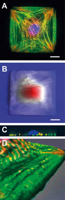 |
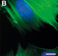
Figure 4: Engineered cardiac myocytes fluorescently labeled for actin (green) α-actinin (red) and DNA (blue) show ordered structure (A). AFM scans of live engineered cells with streaks corresponding to cell beating show a fried-egg like morphology (B) confirmed by confocal microscopy depth projections on fixed cells (C). Zoomed-in detail of 3D-rendered fluorescence and AFM data overlay show that long ridges seen in AFM topography coincide with organized myofibrils (D). Scale bars 10µm. Panels (B) and (C) adapted from Geisse et al. 2009. |
| Understanding the structure and patterning of various organelles in these designer cells and tissues is crucial in this field, and a combined AFM/optical instrument can readily show investigators both the 3D topographical structure as well as the composition of various components within that structure. For example, an epifluorescence ("Sarcomere alignment is regulated by myocyte shape"), confocal, and AFM analysis ("Control of myocyte remodeling in vitro with engineered substrates") of shape-engineered cardiac myocytes showed that the extracellular (out`side) boundary conditions of the cell determine the self-assembly pattern of the intracellular cytoskeleton (Figure 4A,B). Confocal data (Figure 4C) showed that cells segregate the nucleus and cytoskeleton in the vertical direction, and that ridges seen on the AFM topography correspond to the contractile machinery when overlaid onto fluorescence data (Figure 4D). Further, additional studies by Discher and Engler that exploit the force-measuring ability of the AFM have shown that the development of these structures are also influenced by the mechanical properties of the cell-substrate interface ("Myotubes differentiate optimally on substrates with tissue-like stiffness"). | |
| The ability of the AFM to correlate surface topography with internal structural information from fluorescence has also been used to understand mechanisms of membrane fusion in mast cells. Using a combination of topographical mapping and fluorescent staining, Liu and co-workers ("Impact of Actin Rearrangement and Degranulation on the Membrane Structure of Primary Mast Cells: A Combined Atomic Force and Laser Scanning Confocal Microscopy Investigation") were able to identify optically invisible surface membrane ridges that formed concurrently with optically visible F-actin filaments, a previously uncharacterized mechanism. Additionally, it was discovered that the secretory process in these cells is not mediated by actin filaments, suggesting that the cytoskeleton is a poor target for therapeutic strategies. This is important because mast cell degranulation is a key event in allergic and immunoprotective responses. | |
| Expanding on the ability to correlate topography and biochemical information, a combined AFM/ optical system has the added benefit of allowing the investigator to interact with their sample and physically manipulate it. Researchers at UC Berkeley were able to use a carbon nanotube modified AFM tip to penetrate the cell membrane and physically inject molecules of interest without damaging or killing the cell. In this case, transmitted light optical microscopy was used to guide the nano-surgical tool to the area of interest within the cell, while fluorescence microscopy confirmed the internalization, localization, and lifetime of the molecules for several hours after injection into the cell ("A cell nanoinjector based on carbon nanotubes"). This work offers many new possibilities for direct genetic and proteomic manipulation of individual cells, which has been an important strategy in bioengineering, but has been hampered by the relative destructiveness of traditional micron-scale injection techniques. | |
| Electrical and Optical Characterization of Materials | |
| Advances in AFM beyond topographical imaging and force measurement include the characterization of various sample properties including bias, charge, and current flow. One of the most promising future growth areas for the application of these advanced characterization techniques is in the analysis of organic semiconductors and organic photovoltaic materials ("Electrical Scanning Probe Microscopy on Active Organic Electronic Devices"). Fabrication of both device classes results in nanoscale heterogeneities in composition, morphology, and interfaces, all of which can drastically affect functional efficiency. In these cases, electrically-based AFM techniques can provide nanoscale images while simultaneously measuring various electrical properties correlated to topography. This is important for understanding the effects that morphology has on electrical transport properties, which can then be translated to design and fabrication strategies for these devices. | |
  |
|
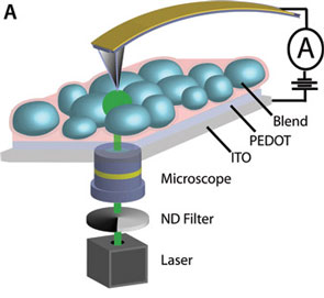
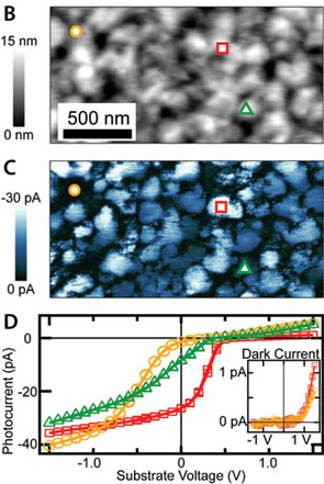
| Figure 5: Optical microscope objectives (A) can be used to stimulate blended organic photovoltaic films, while AFM topography (B) and electrical current (C) maps are recorded. The conductive AFM tip can be placed at specific locations (circle, square, and triangle) for localized current-voltage characterization (D). Reprinted from Coffey et al. 2007. | |
| Ginger and colleagues at the University of Washington have pioneered the application of scanning probe techniques to such organoelectric devices, and their work shows that the combination of these advanced AFM techniques with optical microscopy represents a promising experimental system for their characterization. In one recent study, the investigators measured the nanoscale distribution of light-induced current flow in organic polymer candidates in photovoltaic research ("Mapping local photocurrents in polymer/fullerene solar cells with photoconductive atomic force microscopy"). The technique, known as photoconductive-AFM (pcAFM), uses the AFM to record both the topography and current flow between the tip and the sample, while simultaneously illuminating the sample through the inverted optical microscope objectives. With this system, photocurrent can be correlated with nanoscale topography, and local current-voltage relationships can be measured at individual points across the surface (Figure 5). Further, because of the flexibility of this dual-microscopy approach, illumination intensities across ~8 orders of magnitude could be applied, and the linearity of the intensity-photocurrent relationship was characterized up to several tens of typical solar intensities. | |
| Conclusion and Future Challenges | |
| This is certainly an exciting time in both the development and application of AFM combined with optical imaging techniques. Though there are several studies that exploit the integration of these two imaging modalities beyond those detailed here, the research community has just begun to realize the many possible applications and problems to which these systems can be applied. The broad applicability of the AFM and advanced SPM characterization techniques to a variety of samples across many fields ensures that the field will continue to grow, especially as new challenges arise in multi-disciplinary environments. Coupled to the innovations in optical microscopy developed across centuries, the challenge of designing AFM instrumentation that can be seamlessly integrated with advanced optical techniques while preserving quantitative data collection is paramount to the future success of the technique. Advances and innovations that make current studies possible demonstrate that we are well on our way to a bright future. | |
| By Dr. Nicholas Geisse, Asylum Research | |

Become a Spotlight guest author! Join our large and growing group of guest contributors. Have you just published a scientific paper or have other exciting developments to share with the nanotechnology community? Here is how to publish on nanowerk.com. |
