| Sep 17, 2018 |
2D material produces highest ever signals for human embryonic stem cell detection |
| (Nanowerk Spotlight) For the first time, Singaporean researchers have developed a method using two-dimensional molybdenum disulfide (2D-MoS2) sheets to achieve ultra-high bioelectric signals from human embryonic stem cells (hESCs) using direct current-voltage measurements. |
| This method, which achieved a 1.828 mA cell signal, or two orders of magnitude higher than previous electrical-based detection methods, will pave the way for the development of a broadly applicable, fast, and damage-free stem cell detection method capable of identifying pluripotency with virtually any complementary metal-oxide-semiconductor circuits, the researchers say. |
| "Stem cells are promising starting materials for currently untreated and life-threatening diseases," says Sophia Chan, a PhD Scholar at the Singapore University of Technology and Design. "However, they are limited by readily available methods that can monitor stem cell pluripotency to ensure therapeutic safety. Our method is able to enhance native cell signals feasible for commercialization to ensure therapeutic safety, without altering native cell characteristics." |
| Chan is the first author of a recent ACS Applied Bio Materials paper ("Ultra-High Signal Detection of Human Embryonic Stem Cells Driven by Two-Dimensional Materials") describing the new technique. Her fellow authors are Agency for Science, Technology and Research research fellow Yaw Sing Tan, Nanyang Technological University research fellow Kan-Xing Wu, Nanyang Technological University assistant professor Christine Cheung, and Singapore University of Technology and Design assistant professor Desmond Loke. |
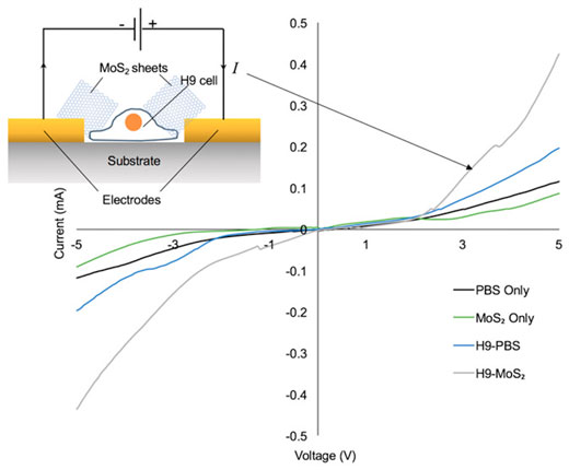 |
| The researchers observed a unique bioelectric signal of human embryonic stem cells using direct current–voltage measurements facilitated by few-layered 2D-MoS2 sheets. A 1.828 mA cell signal was achieved as well as multiple cell reading cycles demonstrating I∼1.9 mA. Native stem cell proliferation, viability, and pluripotency were preserved. Molecular dynamics simulations elucidated the origin of the 2D-MoS2 sheet-assisted increase in current flow. (Reprinted with permission by American Chemical Society) |
Fast, reliable and no damage to cells |
| Electrical-based detection (EBD) methods are excellent candidates for monitoring/validation of pluripotent hESCs. These methods could be used to replace traditional detection methods such as optical-based detection methods to avoid problems such as time-consuming pre-labeling of target proteins with antibodies, and the destruction of differentiated hESCs preventing further cell studies or uses. |
| With past EBD methods however, a difficulty arises from increasing the current flow between the hESCs and metal electrodes while enhancing the cell-electrode adhesion for high device reliability. This has prevented EDB system commercialization. |
| The researchers’ new method overcomes this by utilizing 2D-MoS2 sheets’ good adhesion with metal electrodes and surface molecules unique to stem cells. These materials/molecules essentially allow the current to pass through the cell, 2D-MoS2 sheet, and electrode smoothly, enabling the detection of hESC bioelectric signal using current-voltage measurements. |
| Simulations suggest that the binding between stem cells and 2D-MoS2 sheets is driven by van der Waals interactions and strong electrostatic forces between the cell membrane and the sheet. These interactions assisted the increase in current flow, creating the ultra-high cell signals observed. |
| Enhancing current flow between the cell and metal electrodes would not be useful if the method caused damage to the hESCs or negatively affected the stem cells’ activity. Thus, it was crucial to further understand whether 2D-MoS2 sheets were capable of preserving native stem cell activity. |
| The researchers investigated the dependence of cell morphology, proliferation, and survivability (viability) on incubation time and 2D-MoS2 concentration as well as the pluripotency of stem cells in the presence of 2D-MoS2 sheets. |
| In a time-dependent experiment, stem cells with and without 2D-MoS2 sheets were incubated and fixed for observation at the 24 hour mark and the 48 hour mark. Despite growing at a slightly slower rate, cells incubated with 2D-MoS2 sheets displayed comparable adhesion and cell morphology to the control cells without the sheets. The cells exhibited stable stem cell-like morphologies after 48 hours, indicating that the 2D-MoS2 sheets did not perturb the native cell activity or viability. |
| Comparable to previous studies, high concentrations of 2D-MoS2 sheets appeared to lower cell viability. However, the researchers suggest that using low concentrations of 2D-MoS2 can yield high cell signal values while still maintaining high living stem cell populations. |
| One of the characteristics of hESCs is pluripotency. hESCs are different from normal tissues and organ cells as they can self-renew and be subsequently guided into almost all functional cell lineages. To ensure that the presence of 2D materials were not affecting native pluripotency, cells were incubated with 2D-MoS2 sheets for 24 hours were immunostained for transcription factors essential for maintaining pluripotency. These cells demonstrated fluorescence for the markers, suggesting that the 2D-MoS2 sheets did not affect the pluripotency of the stem cells. |
| "Stem cells are very sensitive compared to other functional cell types. Thus, it was very encouraging to see that the 2D-MoS2 sheets had no significant effects on the sensitive hESCs," Chan says. |
The future of electrical-based detection systems |
| This research will be critical to the development of human embryonic stem cell-based therapy, the researchers say. |
| "Not only can we ensure the safety of future stem cell therapies, we can also deepen our understanding of cell development. This will translate into better and more personalized medication for the future," Chan notes. |
| There is much interest in hESCs as a starting resource to derive functional cells and tissues for regenerative medicine. hESC-based therapy could be used to replace traditional stem cell-based therapies such as transgene-free induced pluripotent stem cell-based therapy to avoid immune rejection. |
| However, clinical applications of hESC-based therapy can only be viable if the monitoring of pluripotent hESCs can be achieved, to minimize the risk of teratoma formation from pluripotent hESCs prior to transplantation for safe clinical uses. |
| "Our method will revolutionize stem cell therapies, getting them one step closer into clinics," Chan hopes. "Not only can it be used to monitor pluripotent stem cells, it can also validate the removal of unwanted pluripotent stem cells prior to transplantation." |
| She says that in principle, this method is applicable to all types of stem cell-detector device structures, 2D materials, and stem cells. Future research could build upon this work to find appropriate combinations of these variables to provide new opportunities for optimizing hESC detection performance. |
|
By Kyra Nian-Yu Wang, Singapore University of Technology and Design
|

Become a Spotlight guest author! Join our large and growing group of guest contributors. Have you just published a scientific paper or have other exciting developments to share with the nanotechnology community? Here is how to publish on nanowerk.com. |
