| Nov 18, 2019 |
Biliverdin nanoparticles pave the way for biodegradable imaging agents |
| (Nanowerk Spotlight) Nanoparticles have shown a lot of promise in biomedical applications such as imaging, drug/gene delivery, and targeted therapy. However, accumulation of nanoparticles in the liver is a major concern, and may be one of the greatest barriers to the widespread adoption of nanoparticles in the clinic. This is especially true for metallic nanoparticles, since the long-term effects of their accumulation in the liver has not been widely studied. |
| A team of researchers from the University of Illinois has looked to nature for inspiration in solving this problem. They decided to use biliverdin, a bile pigment, as the building block for their nanoparticles (ACS Nano, "Biodegradable Biliverdin Nanoparticles for Efficient Photoacoustic Imaging"). |
| "We decided to use biliverdin to make nanoparticles for two reasons: First, biliverdin can naturally be broken down in the liver by an enzyme called biliverdin reductase, which also exists in other organs throughout the body," says Parinaz Fathi, graduate student and first author of this study. "Second, biliverdin has strong absorbance in the near-infrared region, the range of wavelengths in which the body does not have much background absorbance. This allowed us to form nanoparticles that have an inherent capability for as a contrast agent in photoacoustic imaging." |
| Photoacoustic imaging was of particular interest to the team due to its higher depth of penetration and spatial resolution compared to other techniques such as fluorescence imaging. |
| The team took advantage of biliverdin’s multiple carboxylic acid groups and used a small bifunctional molecule as a crosslinker to link multiple biliverdin molecules together. By conducting the nanoparticle synthesis in different liquids, they were able to determine the effects of the synthesis parameters on the nanoparticle behavior. |
| After obtaining biliverdin nanoparticles, the team evaluated their potential for use as a contrast agent. They determined that just like biliverdin, the biliverdin nanoparticles had strong absorbance at wavelengths of 365 nm and 680 nm, with fluorescence and photoacoustic imaging contrast at these wavelengths respectively. |
| They also found that, as expected, biliverdin nanoparticles would degrade after exposure to biliverdin reductase. |
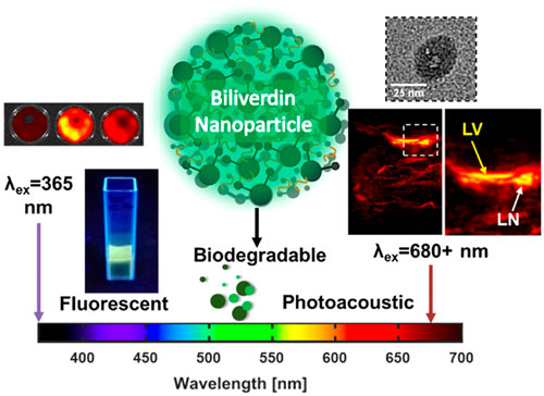 |
| Graphic abstract of this work (© ACS Nano) |
| Having solved the issue of long-term nanoparticle accumulation, the team then looked into the potential for biliverdin nanoparticles to be used in bioimaging. To demonstrate the use of these nanoparticles as photoacoustic contrast agents, the team looked at using them for sentinel lymph node imaging in mice. |
| Sentinel lymph node imaging is especially important because it can allow for the identification of lymph nodes that drain from tumors. Sentinel lymph node biopsies are often conducted to determine how far metastatic cancer has spread throughout the body. |
| The team was able to demonstrate that the biliverdin nanoparticles could be used to identify lymph nodes, and also that the particles would degrade over time within the body. |
| "We were excited to prove the complete degradability of these nanoparticles, which isn’t always possible with the currently reported nanoparticles," said Professor Dipanjan Pan, head of the laboratory for Materials in Medicine. |
|
By Parinaz Fathi, Bioengineering PhD Candidate, NPSC Fellow, NIH T32 TiMe Scholar, University of Illinois at Urbana-Champaign
|

Become a Spotlight guest author! Join our large and growing group of guest contributors. Have you just published a scientific paper or have other exciting developments to share with the nanotechnology community? Here is how to publish on nanowerk.com. |
