| Jan 22, 2021 | |
What is the effect of temperature on a catalytic gold nanoparticle? |
|
| (Nanowerk Spotlight) A team at EMAT, Electron Microscopy for Material Science research group of the University of Antwerp and the NANOlab Center of Excellence, recently investigated the three-dimensional (3D) atomic structure of gold nanoparticles at high temperatures. | |
| The team led by Prof. Sandra Van Aert and Prof. Sara Bals published their work in Nanoscale ("Three-dimensional atomic structure of supported Au nanoparticles at high temperature"). | |
 |
|
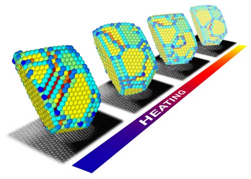
| 3D atomic models of a gold nanoparticle at high temperatures resulting from electron microscopy images acquired from a single viewing direction. (Image courtesy of the researchers) | |
| There is an increasing scientific interest in nanomaterials since they have unique properties that are different from or superior to their bulk counterparts. For example, it is widely known that gold is one of the most stable materials in the world, but when its size reduces to 3-5 nanometres, the gold particles become highly effective catalysts. | |
| Gold nanoparticles are therefore of great importance for a wide range of industrial processes such as CO oxidation, water-gas shift reactions and oxygen reduction at the cathode of hydrogen fuel cells. | |
| It is interesting to note that the properties of gold nanoparticles are directly connected to their size, surface and structure. Hereby, subtle changes in the positions of the atoms at the surface may have a significant impact on their catalytic performance. | |
| Moreover, the surface and structure of these particles are also affected by environmental conditions, such as high temperature or gaseous environments, that are present during catalysis. | |
| Therefore, understanding the structural properties of these nanoparticles at high temperatures is essential to optimize their properties for emerging technologies. | |
| The team at EMAT is a world-leading electron microscopy group with a lot of experience on imaging nanoparticles in 3D. Electron microscopy is an excellent technique to obtain structural information at the atomic-scale, but one typically visualizes a nanoparticle from only one viewing direction. Such a projection image of a 3D particle is insufficient to understand its structure completely. | |
| In their publication, the researchers were able to overcome these limitations by first counting the individual atoms in the particles from high-resolution images. | |
| Next, they used modeling studies to visualize the entire 3D structures of specific gold nanoparticles at high temperatures. The information that was obtained in this study has high scientific importance as it provides insight on how the morphology and the structure of these nanoparticles changes during catalysis. These results may, therefore, help to optimize the design of these materials for novel applications. | |
| Provided by the University of Antwerp | |
|
Become a Spotlight guest author! Join our large and growing group of guest contributors. Have you just published a scientific paper or have other exciting developments to share with the nanotechnology community? Here is how to publish on nanowerk.com. |
|
