| Mar 25, 2021 |
A disposable living laser printed on chip for drug screening |
| (Nanowerk Spotlight) Scientists have created a new way to monitor subtle drug interactions between bacteria and antibiotics. By using a common office inkjet printer, researchers from NTU Singapore and China developed a disposable living laser on chip by encapsulating living bacteria inside. Strong laser emissions generated from bacteria inside the droplet will be dramatically enhanced during drug interactions. This breakthrough could enable more sensitive and high-throughput testing using micro-nano laser technology in the near future. |
| Antibiotics have transformed the field of medicine by making it possible to treat many microbial diseases in nowadays. Monitoring the interactions between bacteria and antibiotics (pathogenic drugs) is therefore a critical step for further evaluation of drug efficacies. Different types of technologies have been developed over the past decade in search for a highly sensitive tool to monitor drug-bacteria interactions. Due to many limitations, conventional techniques usually take longer time to see an obvious result of the drug interactions. It is therefore very challenging to identify tiny dynamic interactions. |
| Recent advances in microlasers have demonstrated its powerful capabilities in terms of signal amplification, strong intensity, and high sensitivity for biomedical sensing. In search for a simpler and more sensitive detection tool, |
| A new study led by Professor Yu-Cheng Chen, Bio+Intelligent Photonics Laboratory, at Nanyang Technological University (NTU Singapore) has now developed a washable, disposable living laser which can monitor tiny dynamic bacteria-drug interactions on a chip. The tiny lasers serve as a highly sensitive culture-free sensor, where living bacteria were encapsulated in the micro-sized water droplets. |
| Excited about their findings published in Analytical Chemistry ("Imaging-Based Optofluidic Biolaser Array Encapsulated with Dynamic Living Organisms"), Prof. Yu-Cheng Chen says, "It’s amazing that these tiny biological living lasers can be directly printed from an office inkjet printer. With the advantages of inkjet printing, the living lasers can be fabricated into a mass dimension in seconds. The cool thing is, you can then wash off the lasers and print again after detection." |
| The preparation of these sensors took place in three steps. First, the researchers labelled the bacteria (Escherichia coli) with nuclei acid dyes, which could recognize the DNA and RNA in cells. Then the cells along with its cell medium were injected into the office printer, where antibiotic drugs can be added directly into the pipet tips (or printhead). Hemisphere micro-droplets were then printed in array on mirror chips. Finally, the living laser array were scanned with a laser beam to generate laser emission images from whispering-gallery modes. |
| As drug interacts with bacteria, the cell membrane would be destroyed and, in turn, more fluorescent DNA (gain molecule) will be released into the droplet through time and contribute to whispering gallery modes, resulting in stronger laser emission. Because laser signal is very sensitive to the changes of the dye molecules at the droplet interface, therefore, tiny increase of the released DNA molecules can be captured and result in a significant change in the gain distribution and laser emissions. |
| The results demonstrated that laser emission image analysis is much more sensitive than fluorescence image analysis by two orders of magnitude, where fluorescence images become saturated after a short period of time. |
| "Our findings show that the amplification that occurs during laser generation enabled us to quantify tiny changes in biological processes in the gain medium," Chen says. |
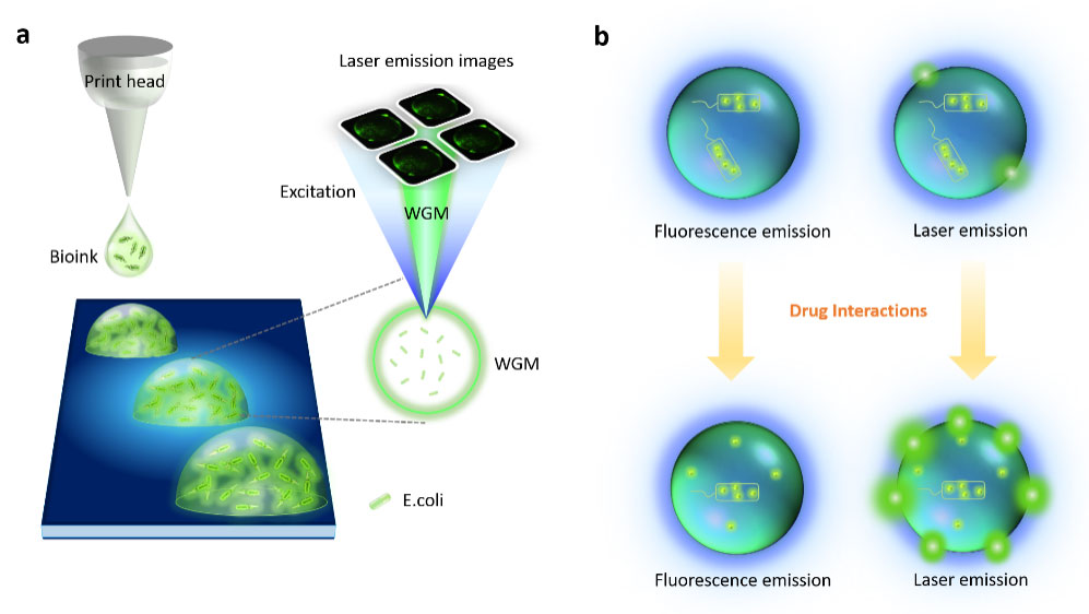 |
| (a) Schematic showing the concept of the image-based E. coli biolaser microarray. (b) Comparison between fluorescence emission and laser emission from a droplet containing E. coli before (top row) and after (bottom row) adding ampicillin. (Reprinted with permission by American Chemical Society) (click on image to enlarge) |
| In collaboration with Shanghai Institute of Microsystem and Information Technology in China, the team's novel living laser biosensor design not only eliminates the need for time-consuming and laborious cell culture, but also simplifies sensor configuration without the requirement of any complex fabrication. |
| Most importantly, a novel concept for bioanalysis based on laser emission images was proposed to quantify the underlying biochemical and biological processes in vitro or in vivo, paving the way for high-throughput on-chip laser analysis of living organisms. |
| Regarding the important future applications of their work, Associate Professor Shilun Feng, who is the co-author explains, "This is a fantastic work by merging microfluidic fabrication with living lasers on a chip. The same approach could be applied to a wide range of living species, including living cells, bacteria, viruses, and proteins interactions. This technology can enable timely diagnosis and treatment with high sensitivity. With the rapid needs for drug screening against viruses, this technology could even enable viruses or bacteria to culture inside the microdroplets and monitor the dynamic interactions with drugs." |
| Indeed, although there is still a long way to fight for many diseases in the future, their device represents a milestone to implement biological living laser towards high-throughput analysis of living organisms. |
| Provided by Nanyang Technological University as a Nanowerk exclusive |

Become a Spotlight guest author! Join our large and growing group of guest contributors. Have you just published a scientific paper or have other exciting developments to share with the nanotechnology community? Here is how to publish on nanowerk.com. |
