Protein Corona
What Is the Protein Corona?
Protein corona The dynamic layer of proteins and other biomolecules that adsorbs onto the surface of a nanoparticle when it enters blood, plasma, serum, lymph, lung lining fluid, cell culture medium, or another biological fluid. The corona often defines the nanoparticle's biological identity: the surface that cells, receptors, complement proteins, and immune cells actually encounter.
The protein corona is one of the central ideas in nanomedicine and nanotoxicology because nanoparticles are rarely seen by the body as the pristine materials prepared in a laboratory. A gold nanoparticle, polymer nanoparticle, iron oxide nanoparticle, silica nanoparticle, or lipid nanoparticle immediately begins to recruit biomolecules from its environment. Within seconds, the engineered surface is partially or completely covered by proteins; over minutes to hours, that coating continues to exchange, reorganize, and mature.
This coating can change the particle's apparent size, surface charge, colloidal stability, receptor engagement, immune recognition, circulation time, tissue distribution, cellular uptake, intracellular fate, and toxicity. In other words, the corona can decide whether a nanoparticle behaves like a long-circulating drug carrier, an immune-system target, a liver-directed delivery vehicle, a diagnostic probe, or a toxic foreign object.
The concept was formalized in influential nanoparticle-protein binding studies in 2007 and later expanded into the idea that biomolecular coronas provide the biological identity of nanosized materials. That framework is now used across biotechnology, biopharmaceuticals, environmental nanoscience, biomaterials, toxicology, and regulatory research.
Synonyms and related terms: nanoparticle protein corona, biomolecular corona, nanoparticle corona, bio-corona, plasma corona, hard corona, soft corona, eco-corona, lipid-protein corona.
Not to be confused with: the coronavirus spike protein or viral corona. In nanomedicine, protein corona refers to a biomolecular coating on a nanoparticle or nanomaterial, not to a virus family.
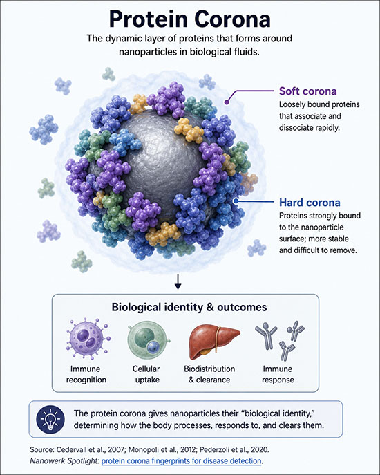
Schematic of the protein corona on a nanoparticle in plasma. A relatively persistent hard corona forms close to the particle surface, while a more weakly associated soft corona exchanges rapidly with surrounding proteins. (Image: Nanowerk)
Why the Protein Corona Matters
Protein corona research matters because it explains a persistent gap between nanoparticle design and biological performance. A particle can be carefully engineered for a target receptor, a desired charge, or a specific biodistribution profile in buffer, yet behave differently in blood. The reason is that cells do not usually interact with the bare synthetic surface. They interact with a protein-decorated surface produced by the local biological environment.
For drug delivery, this can help or hurt. Adsorbed apolipoproteins can direct particles toward hepatocytes; complement and immunoglobulin adsorption can promote phagocytic clearance; albumin adsorption can stabilize some formulations; fibrinogen or denatured proteins can promote inflammatory responses. The corona therefore influences both pharmacology and safety.
For diagnostics, the same phenomenon can be turned into an advantage. A nanoparticle can act as a protein concentrator, selectively enriching a subset of plasma proteins that reflects a person's disease state. Corona-based profiling has therefore become an emerging strategy for biomarker discovery and early disease detection.
How the Protein Corona Forms
Corona formation begins when a nanoparticle enters a protein-containing fluid. Because nanoparticles have high surface area relative to mass, they present many adsorption sites. Proteins diffuse to the surface and bind through hydrophobic interactions, electrostatic attraction, hydrogen bonding, van der Waals forces, metal coordination, and shape complementarity. Binding is affected by both the particle and the surrounding fluid.
The earliest corona is usually influenced by protein abundance. Highly abundant proteins such as albumin, immunoglobulins, fibrinogen, transferrin, and apolipoproteins have many opportunities to collide with the particle. Over time, some early proteins are replaced by proteins with higher surface affinity. This competitive exchange is often described using the Vroman effect, a concept originally developed for protein adsorption on biomaterial surfaces.
The final measured corona is not a fixed universal layer. It depends on particle size, curvature, surface charge, hydrophobicity, roughness, coating chemistry, incubation time, temperature, shear stress, protein concentration, disease state, species, sample handling, and the method used to separate particles from unbound proteins. A corona measured after one hour in fetal bovine serum is not necessarily the same as the corona encountered after intravenous injection in human plasma.
Hard Corona vs. Soft Corona
Researchers often divide the corona into a hard corona and a soft corona. This distinction is useful, but it is also partly operational: it depends on what remains attached after the experimental separation and washing steps.
| Feature | Hard corona | Soft corona |
|---|---|---|
| Binding strength | Relatively strong binding to the nanoparticle surface or to tightly bound proteins | Weaker, more transient association with the particle-corona complex |
| Exchange rate | Slower exchange; residence times can range from minutes to hours or longer | Rapid exchange with proteins in the surrounding fluid, often on a seconds-to-minutes scale |
| Experimental visibility | Easier to isolate by centrifugation, magnetic capture, filtration, or size-exclusion methods | Harder to preserve because it can be lost during washing or dilution |
| Biological relevance | Can dominate longer-lived receptor interactions, uptake pathways, biodistribution, and intracellular fate | Can influence the first moments of cell contact and dynamic interactions in flowing biofluids |
| Common proteins | Apolipoproteins, complement factors, vitronectin, fibrinogen, immunoglobulins, albumin depending on particle type | Exchangeable albumin, transferrin, immunoglobulins, and other weakly associated plasma proteins |
A common pitfall is to assume that the hard corona is the only biologically relevant layer simply because it is easier to measure. In reality, cells can encounter a particle while soft-corona proteins are still present, especially in flowing blood or concentrated tissue fluids. Better methods for preserving or measuring the soft corona are therefore important for making laboratory assays more predictive of in vivo behavior.
What Determines Corona Composition?
Corona composition is determined by a combination of nanoparticle properties, biological environment, and time. Even small changes in formulation can alter the set of proteins recruited to the surface.
| Factor | How it affects the corona | Example consequence |
|---|---|---|
| Particle size and curvature | Curved surfaces expose different contact geometry than flat surfaces; small particles can force proteins into different orientations | Different protein conformations and receptor accessibility |
| Surface charge | Cationic, anionic, and neutral surfaces recruit different protein classes | Cationic particles often show stronger interactions with negatively charged proteins and cell membranes |
| Hydrophobicity | Hydrophobic surfaces tend to bind more proteins and can induce partial unfolding | Higher risk of opsonization, aggregation, or inflammatory signaling |
| Surface coating | PEG, zwitterionic polymers, peptides, sugars, antibodies, and lipids change adsorption patterns | Can reduce nonspecific adsorption or intentionally recruit helpful proteins |
| Biofluid composition | Plasma, serum, lung fluid, cerebrospinal fluid, saliva, and cell culture media contain different protein mixtures | A formulation may behave differently in different tissues or assays |
| Disease state | Cancer, infection, inflammation, pregnancy, metabolic disease, and age can alter the plasma proteome | Patient-specific corona patterns can affect efficacy or serve as diagnostic signatures |
| Time and flow | Early abundant binders can be replaced by higher-affinity proteins; shear stress can alter encounter rates | Static incubation may not match circulation in blood vessels |
Biological Identity and the Nano-Bio Interface
The phrase biological identity captures the key lesson of the corona: the biological system responds to what is present at the nano-bio interface, not merely to what was drawn in the formulation schematic. A targeting ligand, antibody, aptamer, peptide, or sugar on a nanoparticle may be hidden, displaced, denatured, or reoriented after corona formation. Conversely, a corona protein can create a new targeting route that was not intentionally designed.
This is why protein corona research is especially important for active targeting. A nanoparticle decorated with folate, transferrin, RGD peptides, antibodies, or other ligands may show strong binding in protein-free buffer but weaker binding in plasma. Corona proteins can sterically block ligand access, change the apparent charge of the particle, or recruit scavenger receptors and complement receptors that dominate uptake.
The corona also helps explain species differences. A formulation screened in mouse serum may not develop the same corona in human plasma. This can contribute to the weak translation of some nanomedicine results from animal studies to human trials. For clinically intended nanoparticles, corona characterization in relevant human biofluids is therefore more informative than characterization in buffer alone.
Implications for Drug Delivery and Nanomedicine
In drug delivery, the protein corona can influence every major step between injection and therapeutic effect: circulation, aggregation, complement activation, tissue extravasation, receptor binding, endocytosis, endosomal escape, lysosomal trafficking, cargo release, and immune memory. A formulation that appears stable and potent in simple media may lose activity once coated with plasma proteins.
Lipid nanoparticles and RNA delivery
Lipid nanoparticles, the delivery platform used in several RNA medicines and mRNA vaccine technologies, acquire coronas rich in apolipoproteins and other plasma proteins. ApoE adsorption can promote liver uptake through LDL-receptor-related pathways, which is useful for some liver-directed RNA therapies but a barrier for delivery to many other tissues. Recent work has also shown that the LNP corona can affect not only where particles go but also how efficiently mRNA cargo is expressed after cellular uptake.
Stealth coatings and PEGylation
PEGylation is widely used to reduce protein adsorption and extend circulation time. It does not make nanoparticles invisible. PEGylated particles still acquire a corona, often a thinner or compositionally different one. Anti-PEG antibodies, complement activation-related pseudoallergy, and accelerated blood clearance after repeat dosing are important concerns for some PEG-containing nanomedicines.
Opsonization and immune clearance
When immunoglobulins, complement proteins, fibrinogen, or other opsonins bind to a nanoparticle, they can mark it for uptake by macrophages in the liver, spleen, and other parts of the mononuclear phagocyte system. This is one reason many intravenously administered nanoparticles accumulate in the liver and spleen regardless of their intended target. Avoiding harmful opsonization while preserving useful tissue interactions is a central design challenge.
Targeted nanoparticles
Active targeting works only if the targeting moiety remains accessible and functional in the final biological context. Corona formation can mask ligands, alter antibody orientation, or create competing receptor interactions. Successful targeted nanomedicine design therefore requires testing the complete nanoparticle-corona complex, not just the ligand-modified particle in buffer.
The Protein Corona as a Diagnostic Tool
A more recent direction treats the protein corona as a source of information rather than only a delivery obstacle. When a standardized nanoparticle is incubated with plasma from different people, it enriches a subset of proteins from each sample. Because disease alters the abundance, modification, and interaction behavior of plasma proteins, the resulting corona can contain a disease-specific fingerprint.
This approach is attractive because plasma proteomics has a severe dynamic-range problem: a small number of abundant proteins can mask thousands of lower-abundance proteins that may carry diagnostic value. Nanoparticles can act as selective protein concentrators, making otherwise difficult-to-detect signals more visible to mass spectrometry and machine-learning analysis.
Corona-based sensor arrays have been explored for cancer detection and other disease signatures. For a practical example of this emerging diagnostic concept, see Nanowerk's article on protein corona fingerprints for disease detection. The key idea is that no single nanoparticle-protein pattern needs to be universal; a panel of particles with different surface chemistries can generate a richer, disease-informative signature.
Environmental and Toxicology Context
Protein corona research is not limited to injected medicines. Engineered nanomaterials in consumer products, industrial processes, food systems, and the environment encounter proteins, lipids, polysaccharides, natural organic matter, and microbial biomolecules. In these settings, researchers often use the broader term biomolecular corona or eco-corona.
The environmental corona can alter aggregation, dissolution, transport through soil or water, uptake by organisms, and inflammatory effects after inhalation or ingestion. For example, particles contacting lung lining fluid may acquire both lipid and protein layers, producing a surface that differs from the same material suspended in pure water. This matters for occupational exposure, inhalation toxicology, and environmental risk assessment.
How Scientists Measure the Protein Corona
A typical protein-corona experiment incubates nanoparticles in a relevant biofluid, separates the particle-corona complexes from unbound proteins, removes weakly associated background proteins, digests the bound proteins into peptides, and identifies them by liquid chromatography-tandem mass spectrometry. The output is a list of enriched proteins and their relative or absolute abundance.
Mass spectrometry is powerful, but it is not the only tool. Dynamic light scattering and nanoparticle tracking analysis can detect size changes after corona formation. Zeta-potential measurements show changes in apparent surface charge. Isothermal titration calorimetry, surface plasmon resonance, fluorescence correlation spectroscopy, circular dichroism, differential centrifugal sedimentation, electron microscopy, and single-particle methods can help measure binding, conformation, aggregation, and exchange dynamics.
The major experimental challenge is that the act of isolating the nanoparticle can change the corona. Centrifugation can remove soft-corona proteins or force aggregation. Dilution can shift binding equilibria. Washing can erase weak but biologically relevant interactions. For this reason, high-quality corona studies report incubation conditions, separation method, wash stringency, particle recovery, protein controls, and replicate variability.
Typical Research Workflow
A practical corona workflow starts with a biological question: Is this formulation cleared too quickly? Does serum reduce targeting? Does a disease sample create a different corona? Could a specific protein explain toxicity or tissue delivery? Researchers then choose the most relevant biofluid, incubation time, particle concentration, and analytical method.
For a therapeutic nanoparticle, the workflow may compare corona composition in buffer, fetal bovine serum, mouse plasma, and human plasma; test whether major corona proteins change cellular uptake; and validate key proteins by depletion, blocking, or pre-coating experiments. For a diagnostic platform, the workflow may use a panel of nanoparticles, mass spectrometry, statistical feature selection, and blinded validation cohorts.
Strategies to Engineer or Control the Corona
There are three broad strategies for dealing with corona formation. The first is to reduce nonspecific adsorption using hydrophilic or charge-balanced coatings such as PEG, poly(2-oxazoline), zwitterionic polymers, polysarcosine, cell-membrane coatings, or dense carbohydrate layers. This approach aims to limit unwanted opsonization and aggregation, but it rarely eliminates all protein binding.
The second strategy is to pre-form a beneficial corona. Nanoparticles can be coated with albumin, apolipoproteins, dysopsonins, antibodies, peptides, or other proteins before administration. The goal is to create a more predictable interface that improves stability, circulation, tissue delivery, or immune compatibility. Albumin-bound paclitaxel is a clinically familiar example of using a protein-rich nanoparticle interface rather than avoiding proteins altogether.
The third strategy is to design surfaces that recruit a useful corona spontaneously. Instead of treating adsorption as contamination, researchers can tune charge, lipid composition, polymer density, ligand spacing, and curvature to favor proteins that improve delivery. This is especially relevant for RNA delivery, where the best corona may be the one that helps particles reach a tissue and release functional cargo.
Limitations and Common Pitfalls
A major pitfall is assuming that a corona profile is universal. It is not. Corona composition depends on particle formulation, biofluid source, incubation conditions, analytical workflow, and donor biology. A protein list generated under one protocol should not be treated as a fixed identity for that nanoparticle in all contexts.
Another pitfall is overinterpreting abundance. A highly abundant corona protein is not automatically the functionally dominant protein. A lower-abundance protein can control receptor binding, complement activation, or intracellular trafficking if it is exposed in the right orientation or engages a high-affinity receptor.
Cell culture experiments can also be misleading. Serum-supplemented media, serum-free media, and human plasma create different coronas. Fetal bovine serum is convenient but not equivalent to human blood. Protein-free uptake assays may exaggerate targeting performance, while harsh washing steps may remove the soft corona that cells would encounter in vivo.
Finally, corona research must distinguish correlation from mechanism. Finding complement C3, ApoE, albumin, vitronectin, or fibrinogen in a corona suggests possible mechanisms, but functional tests are needed to prove causality. Useful validation experiments include protein depletion, protein add-back, receptor blocking, complement inhibition, orthogonal uptake assays, and animal studies in relevant models.
Key Terms Related to Protein Corona
| Term | Meaning |
|---|---|
| Biological identity | The effective surface identity a nanoparticle presents to cells and tissues after biomolecules adsorb to it |
| Synthetic identity | The designed physicochemical properties of a nanoparticle before exposure to biological fluids |
| Hard corona | Relatively tightly bound proteins that remain associated during isolation and washing |
| Soft corona | Weakly associated, rapidly exchanging proteins surrounding the hard corona |
| Vroman effect | Competitive protein exchange in which early abundant proteins can be replaced by higher-affinity proteins over time |
| Opsonization | Coating by proteins such as immunoglobulins or complement that promotes immune recognition and phagocytic uptake |
| Dysopsonin | A protein that can reduce immune clearance or promote longer circulation |
| Eco-corona | A biomolecular or natural-organic-matter corona formed in environmental settings |
| Proteomics | Large-scale identification and quantification of proteins, often by mass spectrometry |
Frequently Asked Questions
What is a protein corona? A protein corona is the layer of proteins and other biomolecules that adsorbs onto a nanoparticle in blood, plasma, serum, lung fluid, cell culture medium, or another biological fluid. It often becomes the surface that cells and immune systems recognize.
How fast does the protein corona form? Initial adsorption can begin almost immediately after exposure to a biofluid. A measurable corona can form within seconds, but its composition can keep changing over minutes to hours as proteins exchange and reorganize.
Does the corona always make nanoparticles less effective? No. The corona can reduce targeting, increase clearance, or trigger immune reactions, but it can also stabilize particles, direct them to useful receptors, improve liver delivery for some RNA medicines, or provide diagnostic information.
Does PEGylation eliminate the protein corona? No. PEGylation can reduce protein adsorption and prolong circulation, but PEGylated nanoparticles still form coronas. The composition and thickness may change, and anti-PEG immune responses can matter for some applications.
Why are hard and soft coronas difficult to compare between studies? The distinction depends strongly on the isolation method. A harsh centrifugation and wash protocol may leave only tightly bound proteins, while a gentler in situ method may preserve more weakly associated proteins. Reported corona composition is therefore method-dependent.
Can the protein corona be used for disease detection? Yes, in principle. Nanoparticles can enrich disease-informative subsets of the plasma proteome. Multi-particle panels combined with mass spectrometry and statistical analysis are being studied as diagnostic platforms, but large, well-controlled validation cohorts are needed before broad clinical use.
Selected References
Cedervall T., Lynch I., Lindman S., et al. “Understanding the nanoparticle-protein corona using methods to quantify exchange rates and affinities of proteins for nanoparticles.” Proceedings of the National Academy of Sciences 104(7), 2050–2055 (2007). DOI: 10.1073/pnas.0608582104
Monopoli M.P., Åberg C., Salvati A., Dawson K.A. “Biomolecular coronas provide the biological identity of nanosized materials.” Nature Nanotechnology 7, 779–786 (2012). DOI: 10.1038/nnano.2012.207
Walkey C.D., Chan W.C.W. “Understanding and controlling the interaction of nanomaterials with proteins in a physiological environment.” Chemical Society Reviews 41, 2780–2799 (2012). DOI: 10.1039/C1CS15233E
Pederzoli F., Tosi G., Vandelli M.A., Belletti D., Forni F., Ruozi B. “Protein corona and nanoparticles: how can we investigate on?” WIREs Nanomedicine and Nanobiotechnology 9(6), e1467 (2017). DOI: 10.1002/wnan.1467
Voke E., Arral M.L., Squire H.J., et al. “Protein corona formed on lipid nanoparticles compromises delivery efficiency of mRNA cargo.” Nature Communications 16, 9071 (2025). DOI: 10.1038/s41467-025-63726-2