Nanoplasmonics
Nanoplasmonics is a specialized subfield within the broader realm of nanophotonics, as detailed in our comprehensive article on Nanophotonics – harnessing light at the nanoscale. While nanophotonics encompasses the manipulation of light on the nanoscale through various means, nanoplasmonics specifically focuses on the optical phenomena near metal surfaces.
Nanoplasmonics leverages the unique properties of surface plasmons – oscillations of electrons at the surface of metals – to concentrate and manipulate light below the diffraction limit. This aspect of nanophotonics is crucial in applications such as enhancing spectroscopic methods, improving photovoltaic efficiency, and creating sensitive detection systems for biological and chemical molecules
This physical phenomenon of plasmon resonances in small metal particles has been used for centuries. They are visible in the vibrant hues of the great stained-glass windows of the world. Take a prominent historic example: The Lycurgus Cup was created by the Romans in 400 A.D. Made of a dichroic glass – the dichroic effect was achieved by including gold and silver nanoparticles in the glass – the famous cup exhibits different colors depending on whether or not light is passing through it; red when lit from behind and green when lit from in front. It is also the origin of inspiration for contemporary nanoplasmonics research – the study of optical phenomena in the nanoscale vicinity of metal surfaces.

Plasmonic field enhancement
Plasmonic field enhancement is key to numerous applications ranging from surface enhance spectroscopy, sensing, nonlinear optics, light-activated cancer treatments and the enhancement of light absorption in photovoltaics and photocatalysis.
For instance, researchers have developed a tunable nanoantenna that paves the way for new kinds of plasmonic-based optomechanical systems, whereby plasmonic field enhancement can actuate mechanical motion.
Another practical application is a photonic-plasmonic microcavity for ultrasensitive protein detection. This technique optically traps protein molecules at the sites of plasmonic field enhancements in a random gold nanoparticle layer and thus enables the ultrasensite detection of these molecules.
The most intense plasmonic (i.e. optical) fields usually appear within narrow gaps – so-called hotspots – between adjacent metallic nanostructures, especially when the separation goes down to subnanometer scale. However, experimentally probing the plasmonic fields in such a tiny volume still challenges the nanofabrication and detection techniques.
Measuring surface-enhanced Raman scattering (SERS) signal from a probe inside the nanogap region is a promising avenue to do that, but it still faces several intractable issues: 1) how to create a width-controllable subnanometer gap with well-defined geometry; 2) how to insert the nanoprobe into such narrow gap, and more importantly; and 3) how to control the alignment of the probe with respect to the strongest plasmonic field component.
Discovered in the 1970s, SERS is a sensing technique prized for its ability to identify chemical and biological molecules in a wide range of fields – including single-molecule detection sensitivity. It has been commercialized, but not widely, because the materials required to perform the sensing are consumed upon use, relatively expensive and complicated to fabricate. Although the development of a universal substrate for SERS might change that.
SERS relies upon a fundamental phenomenon in physics called the Raman effectthe change in the frequency of monochromatic light (such as a laser) when it passes through a substance. Properly harnessed, Raman scattering can identify specific molecules by detecting their characteristic spectral fingerprints. Potential applications of SERS include the sensing and identification of a range of materials, including chemical and biological agents, improvised explosive devices, and toxic industrial waste.
Raman spectroscopy can provide information about a variety of materials-related phenomena: 1) molecular composition by analyzing bands at characteristic frequencies (fingerprint); 2) symmetry or orientation of molecules or crystals by monitoring peaks using polarization selection for the incident and scattered light; or 3) measurement of stress or strain in a crystal by analyzing e.g., the frequency shift of a characteristic Raman band.
Nanoplasmonics in photovoltaics
Sunlight is composed of many wavelengths of light. In a traditional solar panel, silicon atoms are struck by sunlight and the atoms' outermost electrons absorb energy from some of these wavelengths of sunlight, causing the electrons to get excited. Once the excited electrons absorb enough energy to jump free from the silicon atoms, they can flow independently through the material to produce electricity. This is called the photovoltaic effect – a phenomenon that takes place in a solar panel's photovoltaic cells.
Although silicon-based photovoltaic cells can absorb light wavelengths that fall in the visible spectrum – light that is visible to the human eye – longer wavelengths such as infrared light pass through the silicon. These wavelengths of light pass right through the silicon and never get converted to electricity – and in the case of infrared, they are normally lost as unwanted heat.
Nanoplasmonic techniques that rely on nanostructured metal surfaces could harvest more of the sun's energy.
The use of plasmonic black metals – nanostructured metals are designed to have low reflectivity and high absorption of visible and infrared light – could someday provide a pathway to more efficient photovoltaics (PV) to improve solar energy harvesting.
Plasmonic colors
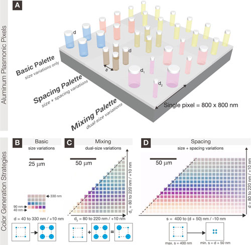
Conventional pigments produce colors by selectively absorbing light of different wavelengths – for example, red ink appears red because it absorbs strongly in the blue and green spectral regions. The reliance on dyes and chemicals for colorants is fraught with problems such as the fading of dyes due to chemical reactions; the need for different dyes for different colors; and the effect of dye-related chemical waste on the environment.
A similar effect can be realized at a much smaller scale by using arrays of metallic nanostructures, since light of certain wavelengths excites collective oscillations of free electrons, known as plasmon resonances, in such structures.
Unlike color pigments which can be overlaid to generate new secondary colors, discrete metal nanostructures rely on size, shape and relative positioning to generate new colors. For instance, aluminum nanostructures can be engineered to produce a broad range of colors, thus making it suitable for ultra-high resolution color printing previously demonstrated in silver and gold. Furthermore, the process of color mixing in nanometallic structures results in 'purer' colors than the basic color palette (read more: "Photorealistic plasmonic printing with aluminum nanostructures").

Nanoplasmonic biosensors
With the potential to revolutionize infection diagnostics, researchers have developed nanoplasmonic sensors capable of detecting live viruses. These label-free optofluidic-nanoplasmonic biosensor have been demonstrated to directly detect live viruses from biological media at medically relevant concentrations with little to no sample preparation.
Another example are simple, ultrasensitive nanoplasmonic microRNA sensors that hold promise for the design of new diagnostic strategies and, potentially, for the prognosis and treatment of pancreatic and other cancers.
And, approaching the nanotechnology holy grail in label-free cancer marker detection – single molecules – researchers have demonstrated the label-free detection of a single protein using a nanoplasmonic-photonic hybrid microcavity.
