Magnetic Tweezers: A Powerful Tool for Manipulating Biomolecules at the Nanoscale
What are Magnetic Tweezers?
Magnetic tweezers are a versatile single-molecule manipulation technique that uses magnetic fields to apply forces and torques on magnetic particles attached to biological molecules, such as DNA, RNA, or proteins. This allows researchers to study the mechanical properties, interactions, and dynamics of these biomolecules at the nanoscale with high precision and control.
Key Components of Magnetic Tweezers
A typical magnetic tweezers setup consists of the following key components:
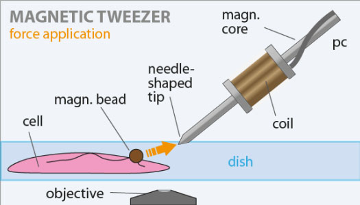
- Magnetic Particles: Superparamagnetic beads, typically made of iron oxide, are attached to the biomolecule of interest. These beads range in size from a few hundred nanometers to several micrometers and can be functionalized with specific chemical groups or antibodies to bind to the target molecule.
- Magnets: Permanent magnets or electromagnets are used to generate a magnetic field gradient that exerts a force on the magnetic particles. The strength and orientation of the magnetic field can be precisely controlled to adjust the applied force and torque.
- Microfluidic Chamber: The sample containing the magnetic particle-tethered biomolecules is placed in a microfluidic chamber, which allows for the exchange of buffers and reagents during experiments. The chamber is typically made of a glass slide and a coverslip separated by a thin spacer.
- Imaging System: A microscope, usually an inverted optical microscope, is used to image the magnetic particles and track their position with nanometer precision. High-speed cameras and image processing algorithms enable real-time tracking of the particles' motion.
Principles of Operation
Magnetic tweezers operate on the principle of applying a force to a magnetic particle attached to a biomolecule. The force is generated by a magnetic field gradient created by the magnets. The magnitude of the force (F) depends on the magnetic moment of the particle (m) and the gradient of the magnetic field (∇B):
F = (m · ∇)B
By controlling the position and strength of the magnets, researchers can precisely tune the force applied to the biomolecule. The force can be used to stretch, twist, or manipulate the molecule, allowing for the study of its mechanical properties, conformational changes, and interactions with other molecules.
The position of the magnetic particle is tracked using video microscopy, providing real-time information about the biomolecule's extension and response to the applied force. By analyzing the particle's motion, researchers can extract quantitative data on the molecule's elasticity, persistence length, and unfolding/refolding dynamics.
Advantages of Magnetic Tweezers
Magnetic tweezers offer several advantages over other single-molecule manipulation techniques, such as optical tweezers and atomic force microscopy:
- Wide Force Range: Magnetic tweezers can apply forces ranging from sub-piconewtons to tens of nanonewtons, making them suitable for studying a wide range of biological processes, from protein unfolding to DNA supercoiling.
- Constant Force Mode: Unlike optical tweezers, which operate in a constant-position mode, magnetic tweezers can maintain a constant force on the biomolecule over extended periods. This allows for the study of time-dependent processes and kinetics.
- Low Photodamage: Magnetic tweezers do not rely on high-intensity laser beams, which can cause photodamage to biological samples. This makes them particularly suitable for studying light-sensitive molecules or processes.
- Torque Application: By using rotating magnets, magnetic tweezers can apply torques to the magnetic particles, enabling the study of DNA supercoiling, protein-DNA interactions, and molecular motors that generate rotational motion.
Applications of Magnetic Tweezers
Magnetic tweezers have found numerous applications in the study of biomolecular mechanics, interactions, and processes at the single-molecule level:
- DNA Mechanics: Magnetic tweezers have been widely used to study the elastic properties of DNA, such as its stretching and twisting behavior. This has provided insights into DNA supercoiling, looping, and packaging in chromatin.
- Protein-DNA Interactions: By tethering DNA molecules between a magnetic bead and a surface, researchers can study the interactions of proteins with DNA, such as the binding and unbinding kinetics of transcription factors, the processivity of DNA-replication enzymes, and the activity of DNA-repair proteins.
- RNA Folding and Dynamics: Magnetic tweezers have been used to investigate the folding and unfolding dynamics of RNA molecules, such as ribozymes and riboswitches. This has shed light on the role of RNA structure in gene regulation and catalysis.
- Molecular Motors: Magnetic tweezers have been employed to study the mechanics and kinetics of molecular motors, such as DNA and RNA polymerases, helicases, and topoisomerases. By applying forces and torques, researchers can probe the stepping behavior, processivity, and energy transduction mechanisms of these enzymes.
The versatility and precision of magnetic tweezers make them a powerful tool for unraveling the mysteries of biomolecular mechanics and interactions at the nanoscale. As the technique continues to evolve, with improvements in sensitivity, throughput, and integration with other methods, it is expected to play an increasingly important role in advancing our understanding of the complex processes that govern life at the molecular level.
Further Reading
Frontiers in Physics, A Guide to Magnetic Tweezers and Their Applications
Annual Review of Biochemistry, High-Resolution Single-Molecule Magnetic Tweezers
